. 2018 May 2;26(5):1181-1197.
doi: 10.1016/j.ymthe.2018.03.003. Epub 2018 Mar 10.

Myeloid Conditioning with c-kit-Targeted CAR-T Cells Enables Donor Stem Cell Engraftment

Affiliations

Myeloid Conditioning with c-kit-Targeted CAR-T Cells Enables Donor Stem Cell Engraftment

Yasuyuki Arai et al. Mol Ther. .

Abstract

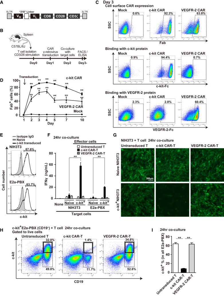
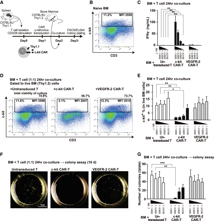
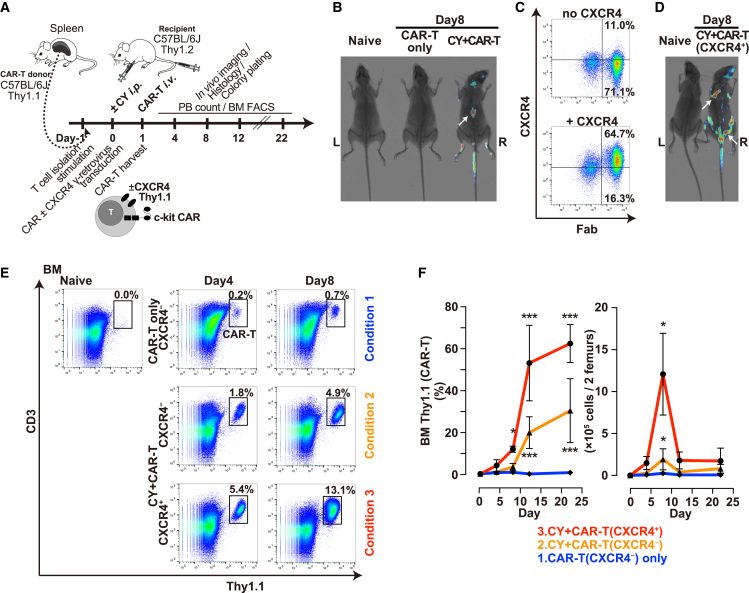
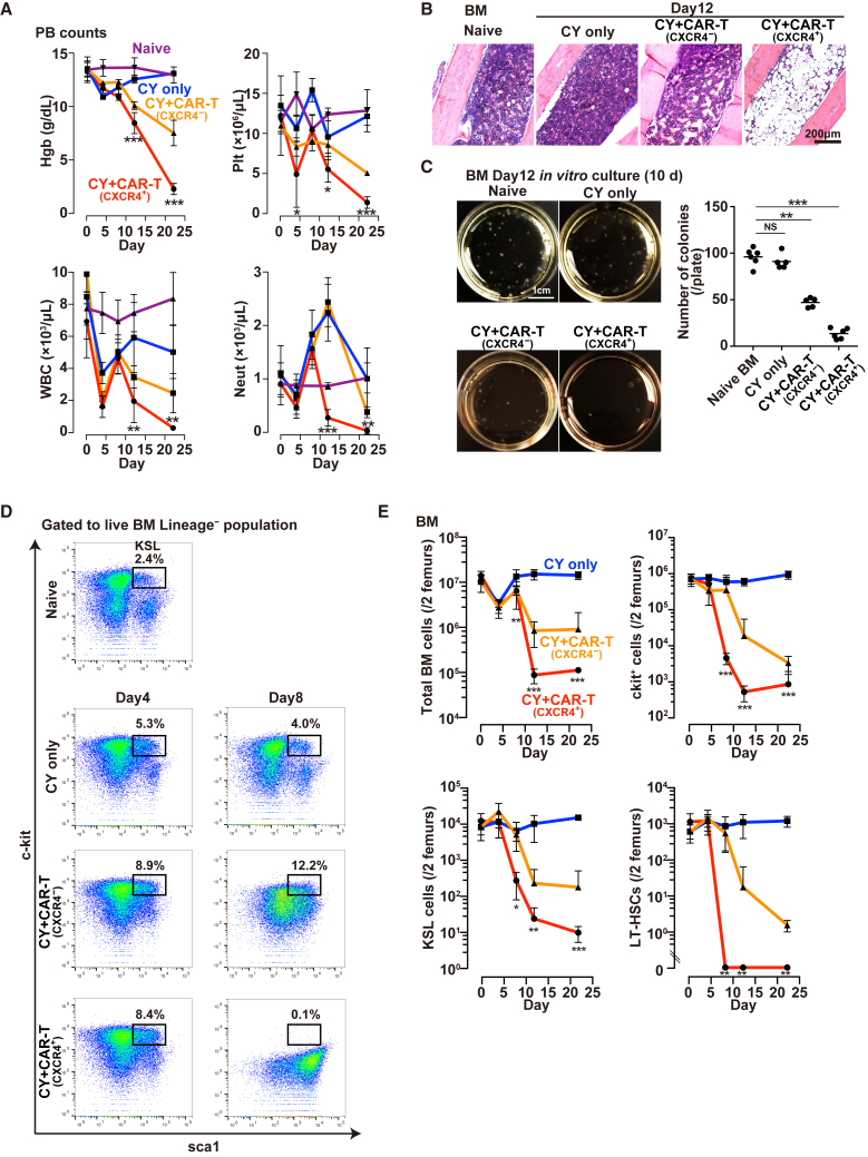
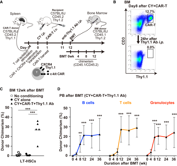
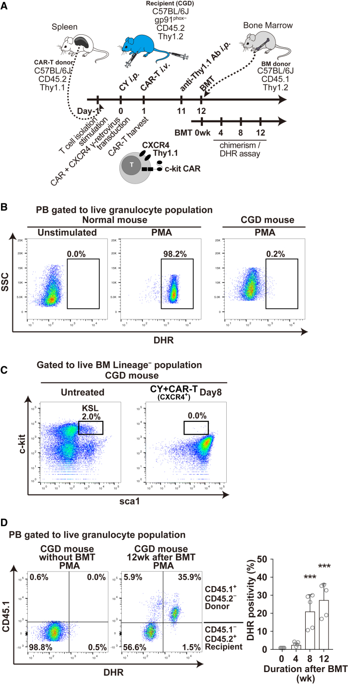
We report a novel approach to bone marrow (BM) conditioning using c-kit-targeted chimeric antigen receptor T (c-kit CAR-T) cells in mice. Previous reports using anti-c-kit or anti-CD45 antibody linked to a toxin such as saporin have been promising. We developed a distinctly different approach using c-kit CAR-T cells. Initial studies demonstrated in vitro killing of hematopoietic stem cells by c-kit CAR-T cells but poor expansion in vivo and poor migration of CAR-T cells into BM. Pre-treatment of recipient mice with low-dose cyclophosphamide (125 mg/kg) together with CXCR4 transduction in the CAR-T cells enhanced trafficking to and expansion in BM (<1%-13.1%). This resulted in significant depletion of the BM c-kit+ population (9.0%-0.1%). Because congenic Thy1.1 CAR-T cells were used in the Thy1.2-recipient mice, anti-Thy1.1 antibody could be used to deplete CAR-T cells in vivo before donor BM transplant. This achieved 20%-40% multilineage engraftment. We applied this conditioning to achieve an average of 28% correction of chronic granulomatous disease mice by wild-type BM transplant. Our findings provide a proof of concept that c-kit CAR-T cells can achieve effective BM conditioning without chemo-/radiotherapy. Our work also demonstrates that co-expression of a trafficking receptor can enhance targeting of CAR-T cells to a designated tissue.

Keywords: CAR-T cells; CXCR4; c-kit; hematopoietic stem cell transplantation; immunotherapy.


Figures

Figure 1
Figure 1
Generation and Evaluation of c-kit CAR-T Cells (A) Schematic map of c-kit CAR γ-retrovirus. (B) Timeline of CAR-T cell production and evaluation. (C) FACS analysis (day 3) of c-kit or VEGFR-2 CAR expression detected by anti-murine Fab (top row). Specific binding of Fc-conjugated murine c-kit or VEGFR-2 with c-kit CAR or VEGFR-2 CAR was detected with anti-Fc fluorescent antibody (middle and bottom rows). (D) Time course of c-kit or VEGFR-2 CAR expression (mean ± SD; n = 5; **p < 0.01 compared with mock-transduced T cells). (E) FACS analysis of c-kit expression on NIH 3T3 or E2a-PBX (CD19+) transduced with c-kit lentivector compared to naive. (F) Generation of IFNγ from c-kit CAR-T cells co-cultured with target lines expressing recombinant murine c-kit (mean ± SD; n = 5; **p < 0.01 comparison as indicated). (G) GFP photomicroscopy images of GFP+ NIH 3T3 (naive or c-kit transduced) co-cultured 24 hr with T or CAR-T cells as indicated after PBS rinse. (H) FACS analysis at 24-hr co-culture assessing cytotoxicity of c-kit CAR-T cells toward c-kit+ E2a-PBX that express CD19. Controls include naive T- or VEGFR-2 CAR-T cells. (I) Percentage of the c-kit+ E2a-PBX within the total E2a-PBX remaining at 24 hr in the cytotoxicity co-cultures, as described for (H) (n = 5; mean ± SD; **p < 0.01).
Figure 2
Figure 2
Congenic Thy1.1 c-kit CAR-T Cell-Mediated Depletion of HSC from Thy1.2 BM In Vitro (A) Timeline of CAR-T production and effect on Thy1.2 BM in vitro. (B) FACS analysis of c-kit+ cells in BM. (C) IFNγ secretion by Thy1.1 T or CAR-T cells in 24-hr co-culture at decreasing cell ratios with Thy1.2 BM (n = 5; mean ± SD; **p < 0.01). (D) FACS analysis assessment of Thy1.1 c-kit CAR-T cell-mediated depletion of c-kit+ Thy1.2 BM cells in 1:1 cell ratio co-culture at 24 hr. Percentge of total cell viability in the culture dishes, percentage of c-kit+ cells in BM population, and their mean fluorescence intensity (MFI) are shown in each panel. (E) Summary results of FACS analyses of the same cytotoxicity co-culture as (D), but at different ratios of T cells to BM (n = 5; mean ± SD; **p < 0.01 and *p < 0.05). (F) Photograph of colony formation assays following Thy1.1 c-kit CAR-T cell-mediated depletion of c-kit+ Thy1.2 BM cells in 1:1 cell ratio co-culture that was plated at 24 hr and read at 10 days. (G) Colony formation numbers in the same experimental conditions and analysis as (F) but at different ratios of T cells to BM (n = 5; mean ± SD; ***p <0.001 and **p < 0.01).
Figure 3
Figure 3
Trafficking and Expansion of Thy1.1 c-kit CAR-T Cells in Thy1.2 Mice (A) Timeline of Thy1.1 CAR-T cell production, in vivo administration, expansion, and trafficking. (B) In vivo whole-body luciferase luminescence imaging overlaid on X-ray image at day 8 following injection of luciferase+ T (or CAR-T) cells with or without CY pre-treatment, where white arrow indicates spleen. Additional mice are shown in Figure S3. (C) FACS analysis of CXCR4 and c-kit CAR on T cells transduced with c-kit CAR alone (upper panel) or co-transduced with CXCR4 (lower panel). (D) In vivo whole-body luciferase luminescence imaging overlaid on X-ray image at day 8 following tail vein injection of luciferase+ CXCR4+ c-kit CAR-T cells into CY-pre-treated mice (right image). White arrows show sites suggestive of BM activity. Additional mice are shown in Figure S3. (E) FACS detection in BM of c-kit CAR or CXCR4+ c-kit CAR-T cells without or with CY pre-treatment as indicated at days 4 and 8 after CAR-T cell administration. (F) Time course of changes in percent (left panel) and actual cell numbers (right panel) of c-kit CAR-T cells in BM using the same conditions as indicated in (E) (n = 5; mean ± SD; ***p <0.001 and *p < 0.05).
Figure 4
Figure 4
In Vivo Depletion of BM Cells after CY Only or after CY + CXCR4+ c-kit CAR-T Cell Treatment (A) Time course of PB hemoglobin (Hgb), platelets (Plt), total leukocyte (WBC), and absolute neutrophil (Neut) count following CY alone (blue), CY + CXCR4 c-kit CAR-T (orange), or CY + CXCR4+ c-kit CAR-T cell (red) compared with the naive (purple) (n = 5; mean ± SD; ***p < 0.001, **p < 0.01, and *p < 0.05). (B) Photomicrographs of H&E-stained tibia sections from naive mouse or mice following CY only, CY + CXCR4 c-kit CAR-T, or CY + CXCR4+ c-kit CAR-T cell injection. (C) Photographs of in vitro colony formation assay of BM cells from naive mice or mice on day 12 following CY only, CY + CXCR4 c-kit CAR-T, or CY + CXCR4+ c-kit CAR-T cell injection. Numbers of colonies per plate are shown in the graph (n = 5; mean ± SD; ***p < 0.001; NS, not significant). (D) FACS analysis of BM obtained from naive mice or from mice at days 4 and 8 after treatment with CY only, CY + CXCR4 c-kit CAR-T, or CY + CXCR4+ c-kit CAR-T cells, where boxes show percentage of KSL (c-kit+sca1+Lineage) cells. (E) Time course of the same FACS analyses of mice as indicated in (D), but calculating and plotting absolute number of BM, c-kit+, KSL, or LT-HSC cells per 2 femurs per mouse (n = 3 [5 mice/experiment; 15 mice total]; mean ± SD; ***p < 0.001, **p < 0.01, and *p < 0.05).
Figure 5
Figure 5
CY Plus Congenic Thy1.1+ CD45.2+ CXCR4+ c-kit CAR-T Cell Facilitated Engraftment of Congenic Donor Thy1.2+ CD45.1+ BM into Thy1.2+ CD45.2+ C57BL/6J Mice following the Depletion of CAR-T Cells with Anti-Thy1.1 Antibody (A) Timeline of c-kit CAR-T cell production and in vivo injection followed by Thy1.1 antibody depletion of c-kit CAR-T cells, transplantation of donor BM, and subsequent evaluation of donor engraftment. (B) FACS analysis of congenic Thy1.1+ CD45.2+ CXCR4+ c-kit CAR-T cells in recipient BM at day 8 comparing before and 24 hr after (day 9) i.p. injection of anti-Thy1.1 antibody (upper versus lower panels). (C) Percentage of all LT-HSCs in recipient BM that are donor phenotype by FACS analysis at 12 weeks after transplant without conditioning, with CY alone, or after CY plus c-kit CAR-T cell conditioning and Thy1.1 antibody depletion of the c-kit CAR-T cells (n = 5; mean ± SD; ***p < 0.001). (D) Time course of percentage of B cell, T cell, and granulocyte lineages in the PB that are donor phenotype by FACS analysis at 0, 4, 8, and 12 weeks and 6 and 9 months after transplant mediated by CY plus c-kit CAR-T cell conditioning and Thy1.1 antibody depletion of the c-kit CAR-T cells (n = 3 [5 mice/experiment; 15 mice total]; mean ± SD; ***p < 0.001 and **p < 0.01).
Figure 6
Figure 6
Treatment of gp91-phox-Deficient CGD Mice with Normal Wild-Type BM Transplantation Using Sequential CY + CAR-T + Thy1.1 Ab Conditioning (A) Timeline of c-kit CAR-T cell production and in vivo injection into CGD mice followed by Thy1.1 antibody depletion of c-kit CAR-T cells, transplantation of donor normal wild-type BM, and subsequent evaluation of donor engraftment and assessment of the appearance of DHR+ oxidase normal granulocyte in the PB of transplanted CGD mice. (B) DHR flow cytometry analysis of ROS production by PB granulocytes without or with PMA stimulation from a normal wild-type mouse (middle and left panels) or CGD mouse (right panel) (gated on granulocyte Gr1+Mac1+ population). (C) FACS analysis of BM obtained from untreated and CY + CAR-T-treated CGD mice at day 8, where boxes show percentage of KSL (c-kit+sca1+Lineage) cells. (D) Normal wild-type donor PB granulocyte chimerism (CD45.1) in c-kit CAR-T cell-conditioned CGD mice (CD45.2) was assessed together with measurement of DHR oxidase activity (PMA stimulation) before transplantation (left panel) and 12 weeks after transplantation of normal wild-type BM into CGD mice conditioned with c-kit CAR-T cells (right panel). Time course of percentage of DHR+ cells (in granulocyte fraction) is shown in right panel at 0, 4, 8, and 12 weeks after transplantation (n = 5; mean ± SD; ***p < 0.001).

Comment in

References

    1. Kang E.M., Marciano B.E., DeRavin S., Zarember K.A., Holland S.M., Malech H.L. Chronic granulomatous disease: overview and hematopoietic stem cell transplantation. J. Allergy Clin. Immunol. 2011;127:1319–1326, quiz 1327–1328. - PMC - PubMed
    1. Heimall J., Puck J., Buckley R., Fleisher T.A., Gennery A.R., Neven B., Slatter M., Haddad E., Notarangelo L.D., Baker K.S. Current Knowledge and Priorities for Future Research in Late Effects after Hematopoietic Stem Cell Transplantation (HCT) for Severe Combined Immunodeficiency Patients: A Consensus Statement from the Second Pediatric Blood and Marrow Transplant Consortium International Conference on Late Effects after Pediatric HCT. Biol. Blood Marrow Transplant. 2017;23:379–387. - PMC - PubMed
    1. Srivastava A., Shaji R.V. Cure for thalassemia major - from allogeneic hematopoietic stem cell transplantation to gene therapy. Haematologica. 2017;102:214–223. - PMC - PubMed
    1. De Ravin S.S., Li L., Wu X., Choi U., Allen C., Koontz S., Lee J., Theobald-Whiting N., Chu J., Garofalo M. CRISPR-Cas9 gene repair of hematopoietic stem cells from patients with X-linked chronic granulomatous disease. Sci. Transl. Med. 2017;9:eaah3480. - PubMed
    1. Czechowicz A., Kraft D., Weissman I.L., Bhattacharya D. Efficient transplantation via antibody-based clearance of hematopoietic stem cell niches. Science. 2007;318:1296–1299. - PMC - PubMed

Publication types