. 2018 Apr 6;360(6384):eaar5078.
doi: 10.1126/science.aar5078.

Triggered recruitment of ESCRT machinery promotes endolysosomal repair

Affiliations

Triggered recruitment of ESCRT machinery promotes endolysosomal repair

Michael L Skowyra et al. Science. .

Abstract

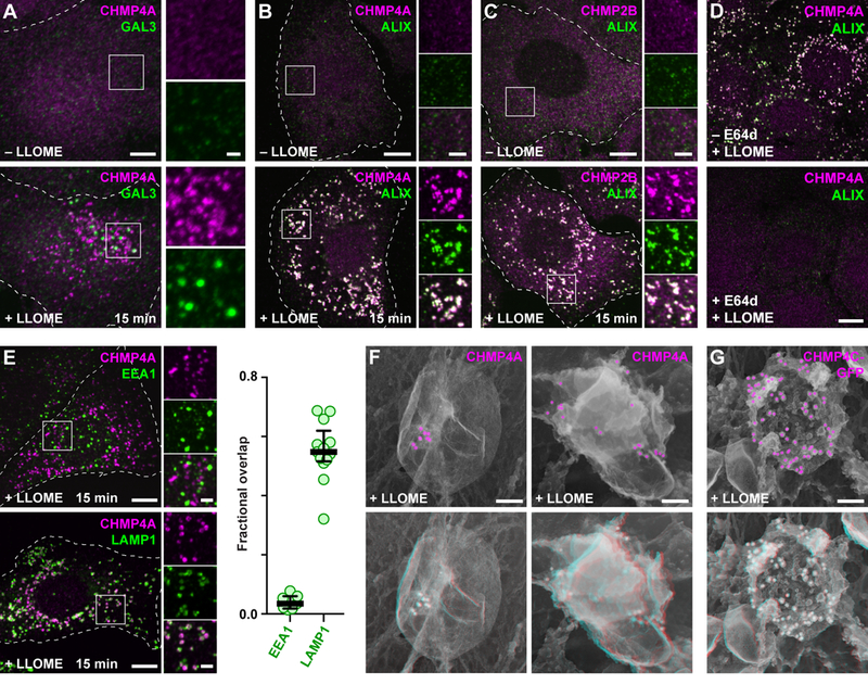
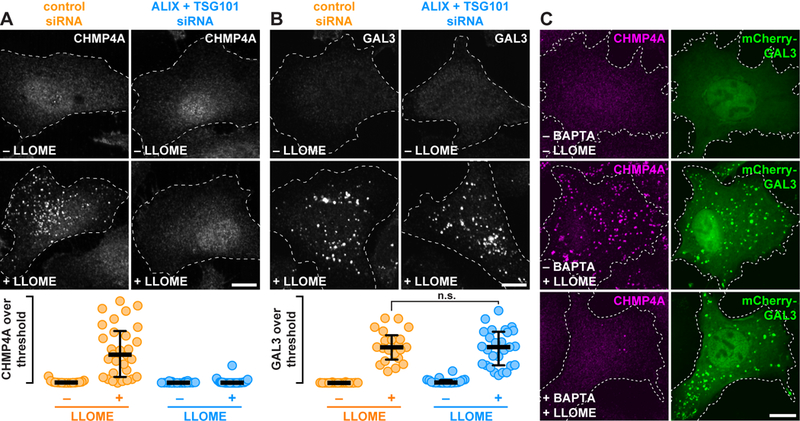
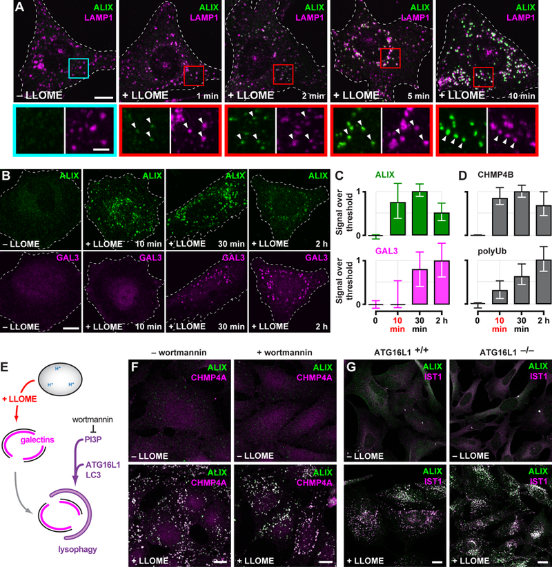
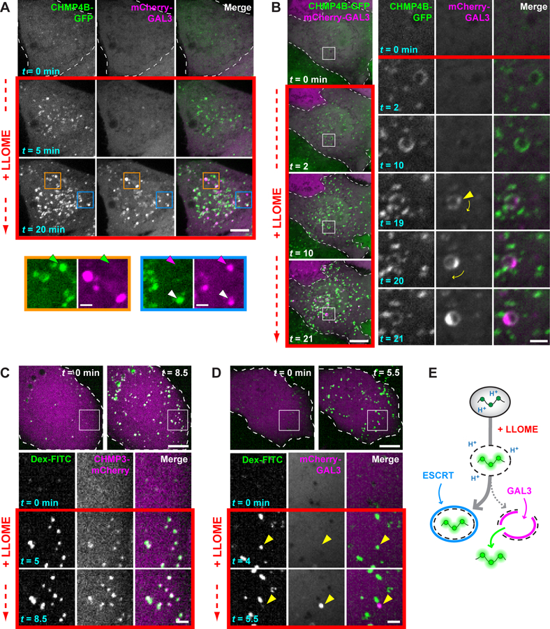
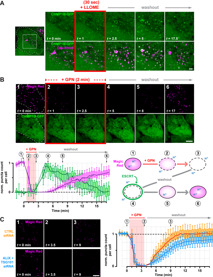
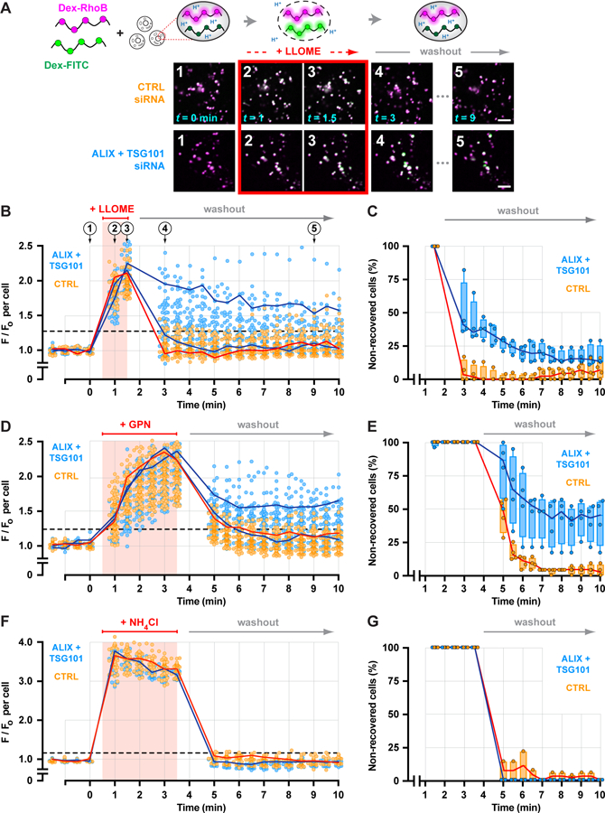
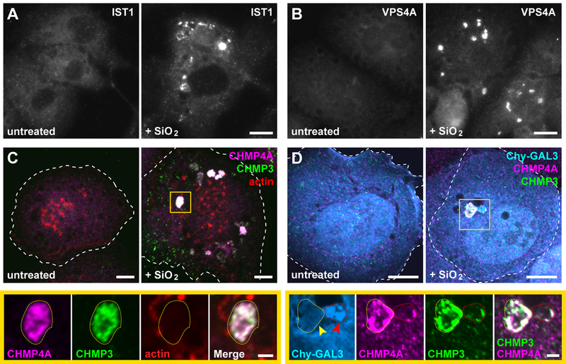
Endolysosomes can be damaged by diverse materials. Terminally damaged compartments are degraded by lysophagy, but pathways that repair salvageable organelles are poorly understood. Here we found that the endosomal sorting complex required for transport (ESCRT) machinery, known to mediate budding and fission on endolysosomes, also plays an essential role in their repair. ESCRTs were rapidly recruited to acutely injured endolysosomes through a pathway requiring calcium and ESCRT-activating factors that was independent of lysophagy. We used live-cell imaging to demonstrate that ESCRTs responded to small perforations in endolysosomal membranes and enabled compartments to recover from limited damage. Silica crystals that disrupted endolysosomes also triggered ESCRT recruitment. ESCRTs thus provide a defense against endolysosomal damage likely to be relevant in physiological and pathological contexts.


Conflict of interest statement

Competing interests: The authors declare no competing financial interests.

Figures

Fig. 1.
Fig. 1.. Damaged endolysosomes recruit ESCRT machinery.
(A) HeLa cells treated with or without LLOME were costained for CHMP4A and GAL3. (B and C) U2OS cells treated as above were costained for ALIX and the indicated ESCRT-III proteins. (D) U2OS cells preincubated for 5 min in the absence or presence of the cathepsin inhibitor E64d were exposed to LLOME and costained for CHMP4A and ALIX. (E) LLOME-treated U2OS cells were stained for CHMP4A and EEA1 or LAMP1. Fractional overlap between CHMP4A puncta and labeled compartments is shown at right (n = 7 cells for EEA1, 13 cells for LAMP1). (F) U2OS cells or (G) HeLa cells producing CHMP4C-GFP were treated with LLOME and immunolabeled as indicated before processing for deep-etch electron microscopy. Top panels depict two-dimensional views with pseudocolored immunogold; bottom panels show corresponding anaglyphs, to be viewed with dual color glasses. In all fluorescence micrographs, representative cells are shown outlined by dashed white lines; boxed areas are magnified at right; and coincidence of green and magenta appears white. Scale bars equal 10 µm (A to E; 2 µm in magnified views); 100 nm (F and G).
Fig. 2.
Fig. 2.. Calcium as well as TSG101 and ALIX are required for ESCRT assembly on damaged endolysosomes.
(A and B) HeLa cells transfected with the indicated siRNAs were treated with or without LLOME and stained for CHMP4A (A) or GAL3 (B). The proportion of each stain that exceeded a starting threshold is quantified (in the order shown: n = 20, 33, 21, and 33 cells in A; and 20, 21, 20, and 27 cells in B; n.s. denotes p = 0.6319 by Student’s two-tailed unpaired t-test). Horizontal bars mark the median; whiskers span the interquartile range. (C) U2OS cells producing mCherry-GAL3 were preincubated for 1 h with or without BAPTA-AM, then treated with DMSO or LLOME and stained for CHMP4A. Scale bars equal 10 µm.
Fig. 3.
Fig. 3.. ESCRT machinery is rapidly recruited to LLOME-damaged endolysosomes and is independent of lysophagy.
(A) U2OS cells exposed to LLOME for the indicated times were costained for ALIX and LAMP1. Boxed areas are magnified below; arrows highlight LLOME-induced ESCRT puncta. (B) HeLa cells treated with LLOME for the indicated times were costained for ALIX and GAL3. The proportion of each stain that exceeded a starting threshold is plotted in (C). Shown are the median and interquartile range (in chronological order: n = 3, 8, 10 and 8 cells). (D) Quantitation of CHMP4B or ubiquitinated material (polyUb) (see fig. S11) as in (C) (in chronological order: n = 10, 11, 15, and 16 cells for CHMP4B; n = 19, 18, 25 and 26 cells for polyUb). (E) Schematic depicting lysophagy and the role of factors targeted in adjacent experiments. (F) U2OS cells preincubated with or without wortmannin were treated with LLOME and costained for ALIX and CHMP4A. (G) HeLa cells expressing endogenous ATG16L1 (ATG16L1+/+) or lacking ATG16L1 as a result of CRISPR-mediated gene deletion (ATG16L1−/−), were treated with or without LLOME for 10 min and costained for ALIX and the ESCRT-III protein IST1. Scale bars equal 10 µm (2 µm in magnified views).
Fig. 4.
Fig. 4.. ESCRT machinery preferentially responds to small membrane disruptions.
(A) HeLa cells producing CHMP4B-GFP were additionally transfected with a plasmid encoding mCherry-GAL3, and imaged live before and after adding LLOME. Colored arrows in magnified views of boxed areas designate structures containing only CHMP4B-GFP (green), only mCherry-GAL3 (magenta), or both (white). (B) Cells imaged as in (A). Boxed areas are magnified at right, with the leftmost two columns showing each signal in grayscale; these are merged and colored in the third column. Arrowhead marks the initial appearance of GAL3. Note that this compartment rotates over time, as denoted by the curved arrows. (C and D) U2OS cells were transfected with plasmids encoding CHMP3-mCherry (C) or mCherry-GAL3 (D) and loaded with 40-kDa pH-sensitive FITC-dextran chased into endolysosomes. Cells were imaged live before and after addition of LLOME. Boxed areas are magnified as in (B). Arrowheads in (D) indicate a dextran-laden compartment that rapidly acquires GAL3 after sudden loss of dextran. (E) Interpretive illustration of the different events seen in (C) and (D). In all panels, single planes of a representative cell are shown at the times indicated from each recording. Scale bars equal 10 µm (2 µm in magnified views).
Fig. 5.
Fig. 5.. Transient ESCRT recruitment accompanies and is important for recovery of endolysosomes from acute damage.
(A) HeLa cells producing CHMP1B-GFP were loaded with 40-kDa Rhodamine B dextran to label endolysosomes, and imaged live during and after a 30 sec exposure to LLOME. Shown are select frames from the resulting timelapse, corresponding to the boxed area of the outlined cell. Similar transient recruitment was observed using GPN (see fig. S23). (B) CHMP1B-GFP cells were incubated with Magic Red to label functional endolysosomes, and then imaged live at 30-sec intervals during and after a 2-min exposure to GPN. Magic Red was maintained at the same concentration throughout (see also Movie S1). The number of Magic Red or CHMP1B-GFP puncta detected at each time is plotted below (n = 17 cells), normalized to the initial number of puncta. Solid lines trace the median; boxes span the interquartile range; error bars designate the 10-th and 90-th percentiles. Dotted lines plot the number of puncta in the cell depicted in the time series, and the corresponding frames are numbered above the graph. An interpretive illustration of the observed events is shown at right. (C) U2OS cells transfected with the indicated siRNA were labeled with Magic Red and recorded live at 30-sec intervals during and after a 3-min pulse of GPN. Left panels depict a representative cell from each cohort, showing dissipation of Magic Red during the GPN pulse and a delay in Magic Red reacquisition in ESCRT-deficient cells. This behavior is plotted at right in terms of the number of Magic Red puncta at each time per cell from each cohort (n = 97 cells from 4 experiments). Scale bars equal 10 µm (2 µm in magnified views).
Fig. 6.
Fig. 6.. Depleting ESCRT machinery impairs endolysosomal membrane repair.
(A) A ratiometric pH-sensing assay was used to monitor endolysosomal integrity. U2OS cells transfected with the indicated siRNAs were loaded with 40–70 kDa dextrans conjugated to pH-sensitive FITC and pH-insensitive Rhodamine B fluorophores chased into endolysosomes. Loaded cells were imaged at 30-sec intervals during and after a 1-min exposure to LLOME. Fluorescence micrographs show a representative field of endolysosomes from each cohort, depicting the changes in FITC fluorescence associated with key steps in the assay. Scale bars equal 10 µm. (B) Changes in the median ratio of FITC-to-Rhodamine B fluorescence per cell at each time are plotted (n = 97 control and 85 knockdown cells from 4 experiments). Red line traces the response of a representative control cell. Blue lines designate two cells from the ESCRT-depleted cohort, one in which recovery was impaired and another in which recovery was similar to control. Horizontal dashed line specifies a recovery threshold derived from the control population (see Methods). In (C), the percentage of cells in each cohort that reached this threshold during recovery was counted, in each of 4 experiments. Lines connect the median; boxes and whiskers span the corresponding quartiles. (D-G) Using the same approach, the behavior of control and ESCRT-deficient cells was compared after a 3-min exposure to GPN (D and E; n = 99 control and 91 knockdown cells from 4 experiments) or to 10 mM ammonium chloride (F and G; n = 43 control and 29 knockdown cells from 4 experiments).
Fig. 7.
Fig. 7.. ESCRTs are recruited to vacuolar compartments disrupted by silica nanocrystals.
(A and B) PMA-differentiated THP-1 monocytes were treated with or without 50 µg/ml silicon dioxide (SiO2) nanoparticles for 3 h and stained for IST1 or VPS4A. Shown are widefield fluorescence images of representative cells from each cohort. (C) THP-1 cells treated as above were co-stained for the indicated ESCRT-III proteins along with rhodamine-conjugated phalloidin. Note that the prominent actin-rich puncta correspond to podosomes characteristic of this cell type. Shown are maximum intensity projections of representative cells. Magnified below are single planes corresponding to the boxed area, highlighting the type of compartment (outlined by yellow dotted lines) on which ESCRTs were predominantly observed in SiO2-fed cells (see fig. S27 for a corresponding three-dimensional rendering). (D) U2OS cells producing mCherry-GAL3 (Chy-GAL3) were fed silica nanoparticles for 1.5 h and costained for the indicated ESCRT-III proteins. Shown are maximum intensity projections and magnified single-plane views of the boxed area. Yellow arrow indicates a compartment (elsewhere encircled by a dotted yellow line) intensely stained for both ESCRT-III proteins but lacking Chy-GAL3; red arrow marks a compartment (elsewhere encircled by a dotted red line) that has accumulated Chy-GAL3 along with some ESCRT. Scale bars equal 10 µm (2 µm in magnified views).

Comment in

  • ESCRTs offer repair service.
    Gutierrez MG, Carlton JG. Gutierrez MG, et al. Science. 2018 Apr 6;360(6384):33-34. doi: 10.1126/science.aat2630. Science. 2018. PMID: 29622643 No abstract available.

References

    1. Paz I et al., Galectin-3, a marker for vacuole lysis by invasive pathogens. Cell Microbiol 12, 530 (2010). - PubMed
    1. Thurston TL, Wandel MP, von Muhlinen N, Foeglein A, Randow F, Galectin 8 targets damaged vesicles for autophagy to defend cells against bacterial invasion. Nature 482, 414 (2012). - PMC - PubMed
    1. Maejima I et al., Autophagy sequesters damaged lysosomes to control lysosomal biogenesis and kidney injury. EMBO J 32, 2336 (2013). - PMC - PubMed
    1. Hung YH, Chen LM, Yang JY, Yang WY, Spatiotemporally controlled induction of autophagy-mediated lysosome turnover. Nat Commun 4, 2111 (2013). - PubMed
    1. Chauhan S et al., TRIMs and Galectins Globally Cooperate and TRIM16 and Galectin-3 Co-direct Autophagy in Endomembrane Damage Homeostasis. Dev Cell 39, 13 (2016). - PMC - PubMed

Publication types

MeSH terms