. 2018 May;7(5):617-629.
doi: 10.1530/EC-18-0067. Epub 2018 Apr 5.

Isolation of a multipotent mesenchymal stem cell-like population from human adrenal cortex

Affiliations

Isolation of a multipotent mesenchymal stem cell-like population from human adrenal cortex

Earn H Gan et al. Endocr Connect. 2018 May.

Abstract

Background: The highly plastic nature of adrenal cortex suggests the presence of adrenocortical stem cells (ACSC), but the exact in vivo identity of ACSC remains elusive. A few studies have demonstrated the differentiation of adipose or bone marrow-derived mesenchymal stem cells (MSC) into steroid-producing cells. We therefore investigated the isolation of multipotent MSC from human adrenal cortex.

Methods: Human adrenals were obtained as discarded surgical material. Single-cell suspensions from human adrenal cortex (n = 3) were cultured onto either complete growth medium (CM) or MSC growth promotion medium (MGPM) in hypoxic condition. Following ex vivo expansion, their multilineage differentiation capacity was evaluated. Phenotype markers were analysed by immunocytochemistry and flow cytometry for cell-surface antigens associated with bone marrow MSCs and adrenocortical-specific phenotype. Expression of mRNAs for pluripotency markers was assessed by q-PCR.

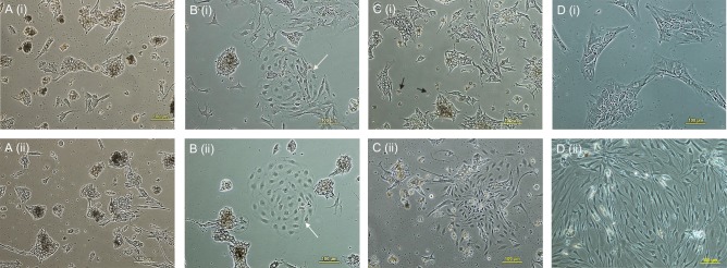
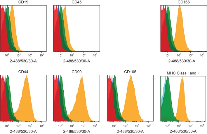
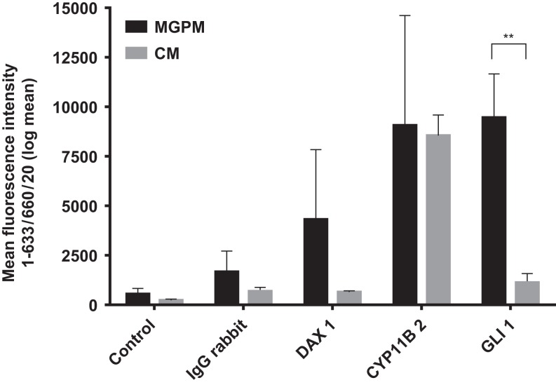
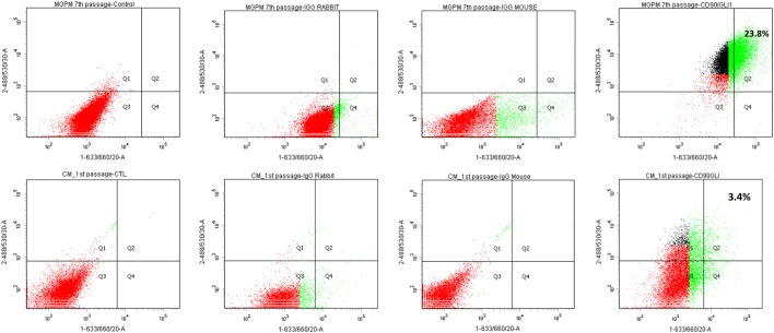
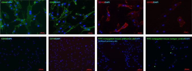
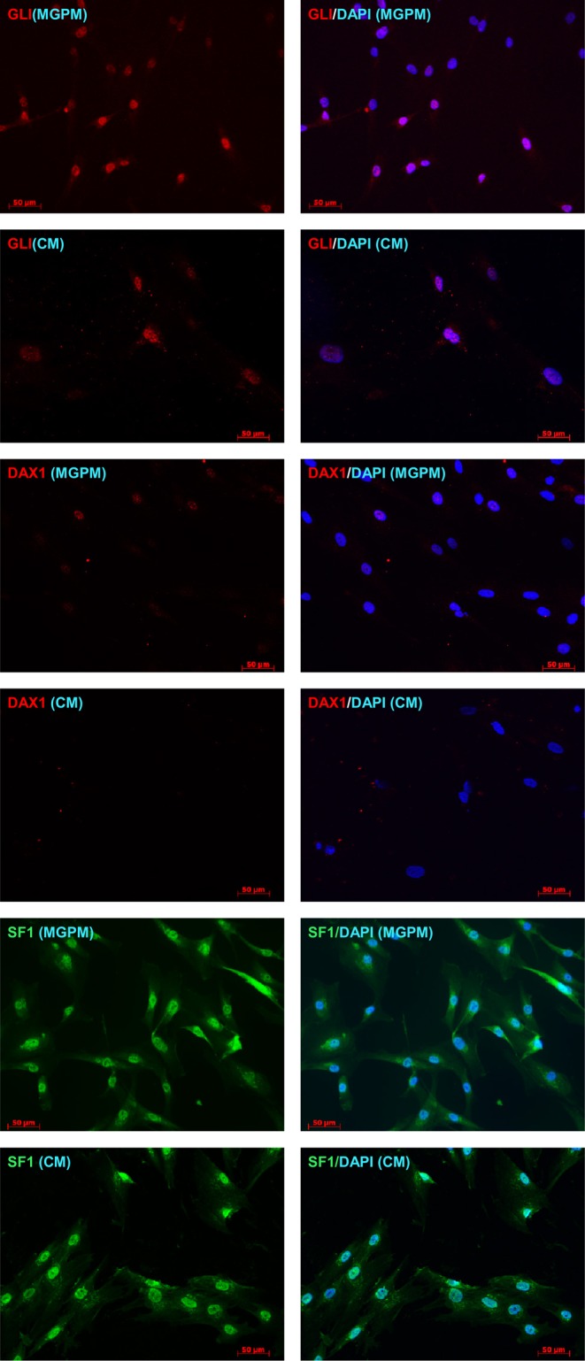
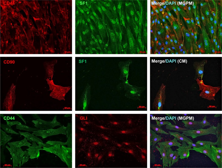
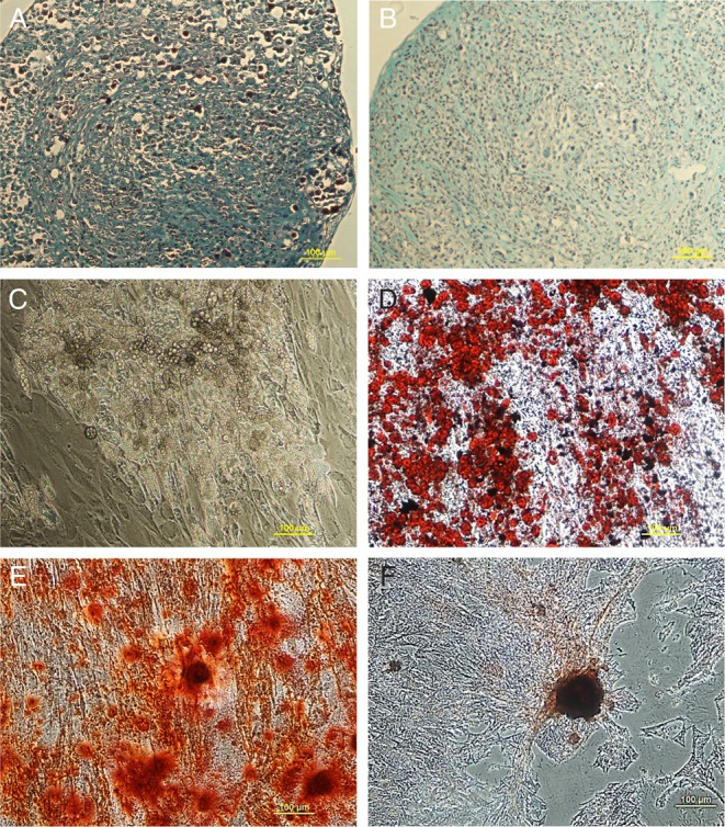
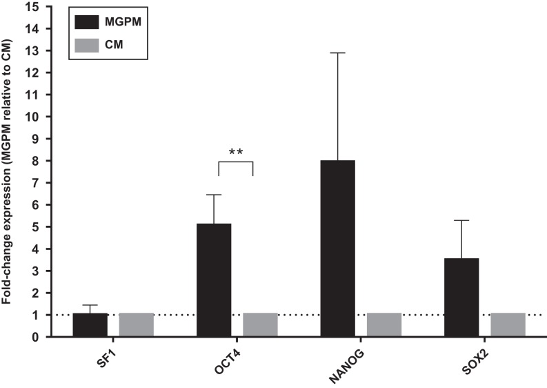
Results: The formation of colony-forming unit fibroblasts comprising adherent cells with fibroblast-like morphology were observed from the monolayer cell culture, in both CM and MGPM. Cells derived from MGPM revealed differentiation towards osteogenic and adipogenic cell lineages. These cells expressed cell-surface MSC markers (CD44, CD90, CD105 and CD166) but did not express the haematopoietic, lymphocytic or HLA-DR markers. Flow cytometry demonstrated significantly higher expression of GLI1 in cell population harvested from MGPM, which were highly proliferative. They also exhibited increased expression of the pluripotency markers.

Conclusion: Our study demonstrates that human adrenal cortex harbours a mesenchymal stem cell-like population. Understanding the cell biology of adrenal cortex- derived MSCs will inform regenerative medicine approaches in autoimmune Addison's disease.

Keywords: adrenocortical stem cell; autoimmune Addison’s disease; mesenchymal stem cells; tissue regeneration.


Figures

Figure 1
Figure 1
Representative images of primary cell cultures seeded in CM (i; upper panel) or MGPM (ii; lower panel), on day 2 (A), day 3 (B), day 5 (C) and passage 3 (D). (A) (i) and (ii) the primary cells seeded in CM or MGPM were predominantly comprised large adrenocortical cells with polygonal appearance and light-refractive core. (B) (i) and (ii) fibroblast-like colony-forming unit (white closed-head arrow) was formed in CM and MGPM respectively. (C) (i) and (ii) primary cells seeded in MGPM were gradually replaced by fibroblast-like cells (ii), whereas adherent cells in CM (i) were predominantly occupied by large polygonal adrenocortical cells with light-refractive lipid core and fragments that protruded from the cells (black closed-head arrow). (D) (i) and (ii) a homogenous cell population comprised fibroblast-like cells continued to thrive in MGPM after third passage (ii). On the contrary, cell population in CM (i) reached senescence following passage 3. (Magnification ×100.)
Figure 2
Figure 2
Representative MSC immunophenotype profile of adrenal cortex-derived cells seeded in CM or MGPM. The panel of histograms represent MSC immunophenotypes of cell populations isolated from human adrenal cortex. The adrenocortical cells displayed strong signals towards CD44, CD90, CD105 and CD166 markers but negative for haematopoitic, lymphocytic markers (CD19 and CD45) and HLA-DR (MHC class II). x-axes display fluorescent signal intensity and y-axes represent cell count. (Red histogram: unstained cells; blue histogram: negative control with fluorochrome-conjugated secondary antibodies only; green histogram: species-specific isotype control; yellow histogram: the staining of cells with antibodies.) The colour codes are applicable to all histograms except graph on MHC class I and II, where blue histogram indicates negative control, yellow and green histograms represent MHC class I (HLA-ABC) and MHC class II (HLA-DR), respectively.
Figure 3
Figure 3
DAX1, GLI1 and CYP11B2 expression among adrenal cortex-derived cell populations cultivated in MGPM vs CM. Cell population harvested in MGPM showed significantly higher GLI1 protein expression (P = 0.046; 95% CI for mean differences 391-16274). DAX1 was expressed in cells cultured in MGPM but there was negligible immunostaining in those harvested from CM (NS). No significant difference was demonstrated in CYP11B2 (expression between the cell populations harvested in either medium. y-axis represents the signal intensity (mean with s.e.m.).
Figure 4
Figure 4
It is a representative dot plot depicting the percentage of cell population (first passage in CM and 7th passage in MGPM) dual-stained with MSC (FITC labelled-488/530/30) and GLI markers (APC-633/660/20). Dual-staining was indicated by the Q2 region, in which >98% of the control and species-specific isotype controls fall outside this area. Approximately 23.8% of the cell population in MGPM co-expressed MSC marker and GLI1 following 7 passages, compared with 3.4% of those cultured in CM after the first passage.
Figure 5
Figure 5
It shows immunolabelling of adrenocortical cells cultured in MGPM for MSC surface markers. Cells from both media showed positive staining for CD 44, CD90, CD105 and CD166 (magnification ×200) but negative staining for CD19 and CD45 markers. (Magnification ×100.)
Figure 6
Figure 6
Adrenocortical cells cultured in MGPM and CM were stained for expression of adrenal-specific (SF1) and stem cell morphogenic signalling markers (GLI1, DAX1) and examined under fluorescence microscopy. Nuclei were stained with DAPI (blue, right hand column). Cells cultured in MGPM showed positive staining for SF1, GLI1 and DAX1. The cell population seeded in CM demonstrated strong SF1 expression but weak GLI with negative DAX1 staining. (Magnification ×200.)
Figure 7
Figure 7
Double-immunostaining of adrenal cortex-derived cells with MSC and SF1/GLI markers. SF1 co-localised with MSC markers in cell populations seeded in either MGPM or CM, as indicated by the membranous staining of CD44/CD90 markers and positive nuclear staining for SF1 markers. Cell population cultured in MGPM also co-expressed MSC and GLI1 marker.
Figure 8
Figure 8
It demonstrates the multilineage differentiation capacity of adrenal cortex-derived cells. Adrenal cells cultured in MGPM were processed into cell pellets and induced with chondrogenic differentiation medium for 21 days. One of three cell pellets (from 3 individual patients) demonstrated weak staining with Safranin O solution, signifying the presence of GAG (A) but two of the remaining 2 showed negative staining (B). Only cells seeded in MGPM survived adipogenic differentiation medium and differentiated into adipogenic cells in abundance, as indicated by cellular accumulation of lipid-rich vacuoles (C), which stained with Oil Red O (D). Cell population seeded in either CM or MGPM (E) or CM (F) differentiated into osteogenic lineage at day-21 with enhancement of alkaline phosphatase activity as shown by the positive staining for Alizarin S.
Figure 9
Figure 9
Adrenal cortex-derived cells cultured in CM and MGPM respectively were analysed by q-PCR for the endogenous expression of pluripotency markers (NANOG, OCT4 and SOX2) and steroidogenic factor 1 marker. Error bars represent standard error of means for cell population from 3 individual patient-derived cultures. Fold changes of gene expression were calculated with the 2−ΔΔCT method, using GUSB as the house keeping gene. Results were shown as mean ± s.e.m. from triplicates (n = 3), using paired T test. Differences in gene expression OCT4 was statistically significant in cell population harvested from MGPM (**P-value <0.05) relative to the cell population cultured in CM.

References

    1. Pearce SH, Mitchell AL, Bennett S, King P, Chandran S, Nag S, Chen S, Smith BR, Issacs JD, Vaidya B. Adrenal steroidogenesis after B lymphocyte depletion therapy in new-onset Addison’s disease. Journal of Clinical Endocrinology and Metabolism 2012. 97 E1927–E1932. ( 10.1210/jc.2012-1680) - DOI - PMC - PubMed
    1. Gan EH, MacArthur K, Mitchell AL, Hughes BA, Perros P, Ball SG, James RA, Quinton R, Chen S, Furmaniak J, et al Residual adrenal function in autoimmune Addison’s disease: improvement after tetracosactide (ACTH1-24) treatment. Journal of Clinical Endocrinology and Metabolism 2014. 99 111–118. ( 10.1210/jc.2013-2449) - DOI - PubMed
    1. Gan EH, Pearce SH. Management of endocrine disease: regenerative therapies in autoimmune Addison’s disease. European Journal of Endocrinology 2017. 176 R123–R135. ( 10.1530/EJE-16-0581) - DOI - PubMed
    1. Ingle DJ, Higgins G. Regeneration of the adrenal gland following enucleation. American Journal of the Medical Sciences 1938. 196 232–239. ( 10.1097/00000441-193808000-00012) - DOI
    1. Zuk PA, Zhu M, Ashjian P, De Ugarte DA, Huang JI, Mizuno H, Alfonso ZC, Fraser JK, Benhaim P, Hedrick MH. Human adipose tissue is a source of multipotent stem cells. Molecular Biology of the Cell 2002. 13 4279–4295. ( 10.1091/mbc.E02-02-0105) - DOI - PMC - PubMed