. 2018 May;7(5):630-636.
doi: 10.1530/EC-18-0116. Epub 2018 Apr 5.

16 kDa vasoinhibin binds to integrin alpha5 beta1 on endothelial cells to induce apoptosis

Affiliations

16 kDa vasoinhibin binds to integrin alpha5 beta1 on endothelial cells to induce apoptosis

Kazunori Morohoshi et al. Endocr Connect. 2018 May.

Abstract

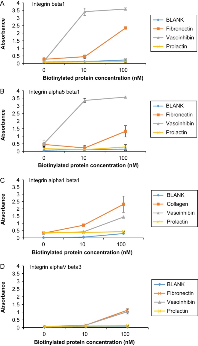
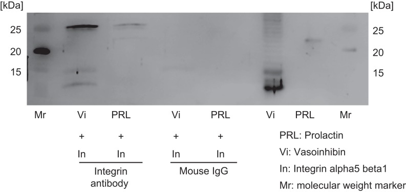
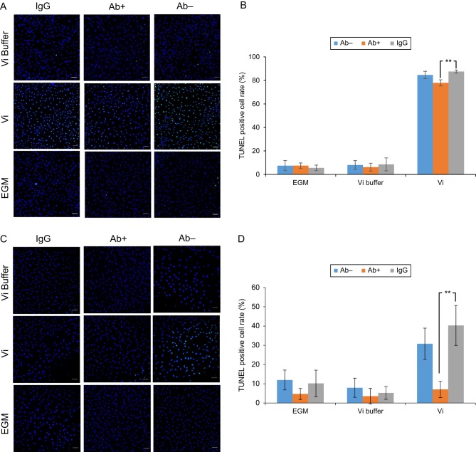
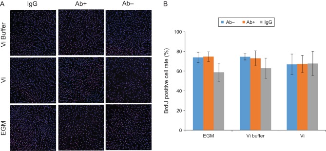
Many functions of vasoinhibins have been reported, but its receptor has not been clarified yet. Vasoinhibins, 11-18 kDa N-terminal fragments of prolactin, have anti-angiogenic activity and act on endothelial cells to induce apoptosis and to inhibit migration and proliferation, which are opposite to the effects of prolactin. Although vasoinhibins bind to the prolactin receptor, its binding activity is very weak compared to prolactin. Therefore, in this study, we evaluated the binding activity between 16 kDa vasoinhibin and integrin beta1, alpha5 beta1, alpha1 beta1 and alphaV beta3 to identify a specific receptor for vasoinhibins. Moreover, we examined whether 16 kDa vasoinhibin induced apoptosis through integrin beta1 and alpha5 beta1 in endothelial cells. In this study, binding assays and co-immunoprecipitation experiments demonstrated that 16 kDa vasoinhibin could bind strongly to integrin beta1 and alpha5 beta1. Moreover, neutralizing with integrin beta1 and alpha5 beta1 antibody could inhibit 16 kDa vasoinhibin-induced apoptosis in endothelial cells. These findings suggest that vasoinhibins can act on endothelial cells through integrin alpha5 beta1 to induce apoptosis.

Keywords: 16 k prolactin; integrin; integrin alpha5 beta1; integrin beta1; vasoinhibins.


Figures

Figure 1
Figure 1
Vasoinhibin binds integrins 0, 10, 100 nM biotinylated proteins were added to 96-well plate coated with integrin beta1 (A), integrin alpha5 beta1 (B), alpha1 beta1 (C) or alphaV beta3 (D) at 100 ng/well concentration. Absorbance was measured 3 h after addition of biotinylated proteins. (A) Absorbance of biotinylated Vi incubated with integrin beta1 was significantly higher than that of biotinylated FN (P < 0.01). (B) Absorbance of biotinylated Vi incubated with integrin alpha5 beta1 was significantly higher than that of biotinylated FN (P < 0.01). (C) Absorbance of biotinylated Vi incubated with integrin alpha1 beta1 was significantly higher than that of BLANK (P < 0.01). (D) Absorbance of biotinylated Vi incubated with integrin alphaV beta3 was significantly higher than that of BLANK (P < 0.01). Data represent mean ± s.d. of 3 independent experiments, double asterisks indicate significant differences (P < 0.01).
Figure 2
Figure 2
Vasoinhibin binds integrin alpha5 beta1 rVi and mPRL after immunoprecipitation of integrin alpha5 beta1 were detected by western blot using an anti-prolactin antibody. Mouse IgG was used for immunoprecipitation as a negative control. rVi was detected after immunoprecipitation, but mPRL was not detected.
Figure 3
Figure 3
Integrin antibody inhibits an apoptotic effect of vasoinhibin incubating HUVEC with Vi for 24 h increases the TUNEL-positive cell rate. Integrin beta1 antibody (A, B) or alpha5 beta1 antibody (C, D) was added to HUVEC before incubation with Vi. (A and C) Immunofluorescence images showed that the integrin antibodies decreased TUNEL-stained cell (green). Nuclei are stained blue. Scale bar indicates 100 µm. (B) Integrin beta1 antibody decreased the TUNEL-positive cell rate induced by Vi (P < 0.01). (D) Integrin alpha5 beta1 antibody also decreased TUNEL-positive cell rate induced by Vi (P < 0.01). Data represent mean ± s.d. of 3 independent experiments, double asterisks indicate significant differences (P < 0.01).
Figure 4
Figure 4
Incubating HUVEC with Vi for 24 h did not affect the BrdU-positive cell rate. Integrin alpha5 beta1 antibody was added to HUVEC before incubation with Vi. (A) BrdU-positive cells were identified by immunohistochemistry (red). Nuclei are stained blue. Scale bar indicates 100 µm. (B) Vi did not affect the BrdU-positive cell rate in this experimental condition. Data represent mean ± s.d. of 3 independent experiments.

References

    1. Cooke NE, Coit D, Shine J, Baxter JD, Martial JA. Human-prolactin – cDNA structural-analysis and evolutionary comparisons. Journal of Biological Chemistry 1981. 256 4007–4016. - PubMed
    1. Devos AM, Ultsch M, Kossiakoff AA. Human growth-hormone and extracellular domain of its receptor – crystal-structure of the complex. Science 1992. 255 306–312. ( 10.1126/science.1549776) - DOI - PubMed
    1. Struman I, Bentzien F, Lee HY, Mainfroid V, D'Angelo G, Goffin V, Weiner RI, Martial JA. Opposing actions of intact and N-terminal fragments of the human prolactin growth hormone family members on angiogenesis: an efficient mechanism for the regulation of angiogenesis. PNAS 1999. 96 1246–1251. ( 10.1073/pnas.96.4.1246) - DOI - PMC - PubMed
    1. Clapp C, Aranda J, Gonzalez C, Jeziorski MC, de la Escalera GM. Vasoinhibins: endogenous regulators of angiogenesis and vascular function. Trends in Endocrinology and Metabolism 2006. 5 301–307. ( 10.1016/j.tem.2006.08.002) - DOI - PubMed
    1. Piwnica D, Touraine P, Struman I, Tabruyn S, Bolbach G, Clapp C, Martial JA, Kelly PA, Goffin V. Cathepsin D processes human prolactin into multiple 16K-like N-terminal fragments: study of their antiangiogenic properties and physiological relevance. Molecular Endocrinology 2004. 18 2522–2542. ( 10.1210/me.2004-0200) - DOI - PubMed

LinkOut - more resources