. 2018 Feb 27;51(1):9-20.
doi: 10.1267/ahc.17020. Epub 2018 Feb 21.

Improved Serial Sectioning Techniques for Correlative Light-Electron Microscopy Mapping of Human Langerhans Islets

Affiliations

Improved Serial Sectioning Techniques for Correlative Light-Electron Microscopy Mapping of Human Langerhans Islets

Sei Saitoh et al. Acta Histochem Cytochem. .

Abstract

Combined analysis of immunostaining for various biological molecules coupled with investigations of ultrastructural features of individual cells is a powerful approach for studies of cellular functions in normal and pathological conditions. However, weak antigenicity of tissues fixed by conventional methods poses a problem for immunoassays. This study introduces a method of correlative light and electron microscopy imaging of the same endocrine cells of compact and diffuse islets from human pancreatic tissue specimens. The method utilizes serial sections obtained from Epon-embedded specimens fixed with glutaraldehyde and osmium tetroxide. Double-immunofluorescence staining of thick Epon sections for endocrine hormones (insulin and glucagon) and regenerating islet-derived gene 1 α (REG1α) was performed following the removal of Epoxy resin with sodium ethoxide, antigen retrieval by autoclaving, and de-osmification treatment with hydrogen peroxide. The immunofluorescence images of endocrine cells were superimposed with the electron microscopy images of the same cells obtained from serial ultrathin sections. Immunofluorescence images showed well-preserved secretory granules in endocrine cells, whereas electron microscopy observations demonstrated corresponding secretory granules and intracellular organelles in the same cells. In conclusion, the correlative imaging approach developed by us may be useful for examining ultrastructural features in combination with immunolocalisation of endocrine hormones in the same human pancreatic islets.

Keywords: Epon-embedded; compact and diffuse islets; correlative microscopy; hormone; immunostaining.


Figures

Fig. 1.
Fig. 1.
Schematic representation of serial sectioning techniques for correlative light-electron microscopy mapping of human Langerhans islets. A: Schematic images illustrating an ultrathin section of a Langerhans islet mounted on a Φ1 × 2 mm single slit copper grid with a formvar film covered by evaporated carbon. B: Flow chart of pre-treatments and immunohistochemical staining procedures applied to serial thick sections after ultrathin sectioning of Epon blocks.
Fig. 2.
Fig. 2.
Differences between compact and diffuse islets in human pancreas revealed by light microscopy observations of toluidine blue stained thick sections routinely embedded in Epon. A: The compact islet appears round to oval and is composed of endocrine cells surrounded with a capsule of connective tissue (black arrowheads). Blood capillaries (black arrows) separate the islet into several lobules. Endocrine cells have moderately light cytoplasm. B: The diffuse islet is composed of a mass of endocrine cells interspersed between adjacent exocrine acinar-like cell clusters without a clear capsule (red arrowheads) in structures that appear like acinar ducts. Bars = 20 μm.
Fig. 3.
Fig. 3.
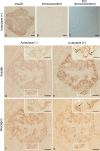
Enhancement of immunostaining for insulin and glucagon in serial thick sections of a typical compact human pancreatic islet by autoclave treatment. A: Insulin-positive staining in a section that underwent autoclave treatment (AC). B: Immunocontrol incubated with secondary antibody without the primary antibody. C: Differential interference contrast microscope (DIC) image of the immunocontrol section. D: Weak insulin immunoreactivity in an untreated section. E: Stronger insulin immunoreactivity in a section that underwent AC—tiny granular patterns are more clearly detected throughout the cytoplasm of β cells (inset, arrows). F: Improved immunoreactivity for glucagon in the cytoplasm of cells in an untreated section. G: Clear granular pattern of immunoreactivity for glucagon in a section that underwent AC (inset, arrows). Bars = 20 μm (A–G); 10 μm (D–G, insets).
Fig. 4.
Fig. 4.
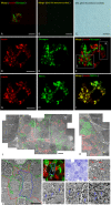
Correlative light-electron microscopy mapping of a compact islet using serial Epon sections from a human pancreatic tissue specimen. Ultrastructural observations and double-immunofluorescence staining for insulin, glucagon, and REG1α were carried out. A: Merged image of insulin and glucagon positive staining. B: Merged image of immunocontrol (red and green) exposed to two secondary antibodies without primary antibodies. C: Differential interference contrast microscope (DIC) image of immunocontrol section shown in (B). Islet cells with granular immunopositive staining for insulin (A, D, F, G, I, red) are immunopositive for REG1α (H, I, green), but not for glucagon (A, E, F, green), whereas the immunostaining patterns of insulin and REG1α largely overlap (I, yellow). Electron microscopic montages (J, K) obtained from the same field of the serial section shown in (F). The magnified image (J, K) corresponds to the white boxes in (F). The compact islet has a capsule of connective tissue (J, K, yellow asterisks). Mapping images of β cell granules (J, K, pseudo-coloured red) and α cell granules (J, K, pseudo-coloured green) manually coloured by Photoshop. Distribution patterns of insulin- (red) and glucagon-positive (green) cells (A, D–F) are clearly identified in the electron micrographs (J, K). Highly magnified electron microscopy images (O–R) correspond to the white boxes in (L) and double-immunopositive staining images (M). N: Toluidine blue staining for the same section (F) of insulin-positive cells (M, red) included β-cell granules (O, red arrows) and condensing vacuoles (O, white arrows), while glucagon-positive cells (M, green) included small numbers of round-shaped granules with low (P, blue arrows), with intermediate (Q, blue arrows) and high (R, blue arrows) electron densities. Bars = 20 μm (A–D, G); 1 μm (J–L).
Fig. 5.
Fig. 5.
Different distribution of HRP-DAB-immunoreaction products for REG1α and insulin in the same endocrine cells of diffuse type islet in serial thick sections of a diffuse human pancreatic islet. The diffuse islet is composed of a mass of endocrine cells interspersed between adjacent exocrine acinar-like cell clusters without a clear capsule (A, arrowheads). Islet endocrine cells show granular immunopositive staining for REG1α (B) or insulin (C), while acinar-like cells in contact with islet endocrine cells were not immunostained. TB: toluidine blue staining. Bars = 20 μm.
Fig. 6.
Fig. 6.
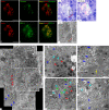
Correlative light-electron microscopy mapping of a diffuse islet using serial Epon sections from a human pancreatic tissue specimen. Ultrastructural observation and double-immunofluorescence staining for insulin, glucagon, and REG1α were carried out. Granular immunoreactivities for insulin (A, C, red) and glucagon (B, C, green) are sparse and dense respectively, while the immunostaining patterns of insulin (D, F, red) and REG1α (E, F, green) largely overlap. G: A light microscopy image of the same thick section stained with toluidine blue (TB) after microscopy observation of fluorescence immunostaining (A–C). The diffuse islet is composed of a mass of endocrine cells in contact with adjacent exocrine acinar-like cell clusters without a clear capsule (red arrowheads). H: Superimposed image of (C) and (G). I: An electron microscopy image of the black-boxed area shown in (G) obtained from a serial ultrathin section demonstrating the ultrastructural features of insulin- and glucagon-positive endocrine cells in a diffuse pancreatic islet. The diffuse islet is composed of a mass of endocrine cells interspersed between adjacent exocrine acinar-like cell clusters without a clear capsule (red arrowheads). J: An electron microscopy montage showing a pancreatic islet corresponding to the rectangle of TB staining shown in (G). Endoplasmic reticulum and Golgi apparatus are indicated by blue and red arrowheads, respectively. K: A higher magnification image of the respective boxed area in (J) illustrating an α cell containing α-cell granules (red arrowheads) in contact with a β cell containing β-cell granules (blue arrowheads) and condensing small vacuoles (cyan arrowheads). L: A higher magnification image of the respective boxed area in (J) illustrating a β cell with β-cell granules (blue arrowheads) and spherical and smaller granules with small halo (green arrowheads) in contact with an α cell containing α-cell granules (red arrowheads). M: A higher magnification image of the respective boxed area in (J) illustrating a β cell with β-cell granules (blue arrowheads) and a zymogen-like granule (yellow arrow) in cell-to-cell contact with an exocrine acinar-like cell containing zymogen-like granules. N: Interdigitation of cell membranes (white arrows) containing β-cell granules (blue arrowheads) and 200–500-nm condensing vacuoles (cyan arrowheads) between two β cells. Bars = 50 μm (A, D, G); 5 μm (I); 2 μm (J); 500 nm (K, N); 1 μm (L, M).
Fig. 7.
Fig. 7.
Serial block-face scanning electron microscopy (SBF-SEM) observations of compact and diffuse islets from pancreatic tissues of a C57BL/6J mouse. A: The compact islet is composed of a mass of endocrine cells with a clear capsule from the tail portion of the pancreas. B: α cells and β cells localized in the corner of an islet in contact with a capsule. C: α-cell granules (blue arrowheads) are electron dense without a core, while β-cell granules (red arrowheads) show angular-shaped cores with a clear halo. D–E: The diffuse islet is composed of a mass of endocrine cells in contact with adjacent exocrine acinar-like cell clusters (ATLANTIS) (red arrowheads) without a clear capsule (blue arrowheads). F: The islet endocrine cells contain three type of granules: 1) spherical smaller granules with a small halo (green arrowheads), 2) slightly electron opaque but spherical granules without a halo having a similar size to α-cell granules (cyan arrowheads), and 3) granules with zymogen-like granules (yellow arrows). The ATLANTIS containing β-cell granules (red arrowheads). G: Montage picture of the islet endocrine cells and ATLANTIS in the diffuse islet (D) showing every 10 serial images of a total of 70 images at 50-nm thickness each. The ATLANTIS containing zymogen-like granules (G10th, white arrows), isotopically localized to the islet cells (G10th, blue arrows), and in direct contact with endocrine cells with lamellar endoplasmic reticuli (G10th, red arrowheads). Islet cells are partially separated from the acinar-like cell clusters with connective tissues by blood vessels (G70th, blue arrowheads). H: Magnified image of (G10th). I: Three-dimensional reconstruction of serial images indicating the zymogen-like granules (shown in orange) inside the islet endocrine cells in contact with ATLANTIS.

Similar articles

Cited by

References

    1. Aida K., Saitoh S., Nishida Y., Yokota S., Ohno S., Mao X., Akiyama D., Tanaka S., Awata T., Shimada A., Oikawa Y., Shimura H., Furuya F., Takizawa S., Ichijo M., Ichijo S., Itakura J., Fujii H., Hashiguchi A., Takasawa S., Endo T. and Kobayashi T. (2014) Distinct cell clusters touching islet cells induce islet cell replication in association with over-expression of Regenerating Gene (REG) protein in fulminant type 1 diabetes. PLoS One 9; e95110. - PMC - PubMed
    1. Baskin D. G., Erlandsen S. L. and Parsons J. A. (1979) Immunocytochemistry with osmium-feed tissue. 1. Light microscopic localization of growth hormone and prolactin with the unlabeled antibody-enzyme method. J. Histochem. Cytochem. 27; 867–872. - PubMed
    1. Bendayan M. and Zollinger M. (1983) Ultrastructural localization of antigenic sites on osmium-fixed tissues applying the protein A-gold technique. J. Histochem. Cytochem. 31; 101–109. - PubMed
    1. Ben-Othman N., Vieira A., Courtney M., Record F., Gjernes E., Avolio F., Hadzic B., Druelle N., Napolitano T., Navarro-Sanz S., Silvano S., Al-Hasani K., Pfeifer A., Lacas-Gervais S., Leuckx G., Marroquí L., Thévenet J., Madsen O. D., Eizirik D. L., Heimberg H., Kerr-Conte J., Pattou F., Mansouri A. and Collombat P. (2017) Long-term GABA administration induces alpha cell-mediated beta-like cell neogenesis. Cell 168; 73–85. - PubMed
    1. Bensley R. R. (1911) Studies on the pancreas of the guinea pig. Am. J. Anat. 12; 297–388.