. 2017 Jun 20;27(23):1700995.
doi: 10.1002/adfm.201700995. Epub 2017 Apr 24.

Multifunctional biomedical imaging in physiological and pathological conditions using a NIR-II probe

Affiliations

Multifunctional biomedical imaging in physiological and pathological conditions using a NIR-II probe

Kangquan Shou et al. Adv Funct Mater. .

Abstract

Compared with imaging in the visible (400 - 650 nm) and near-infrared window I (NIR-I, 650 - 900 nm) regions, imaging in near-infrared window II (NIR-II, 1,000-1,700 nm) is a highly promising in vivo imaging modality with improved resolution and deeper tissue penetration. In this work, a small molecule NIR-II dye,5,5'-(1H,5H-benzo[1,2-c:4,5-c'] bis[1,2,5]thiadiazole)-4,8-diyl)bis(N,N-bis(4-(3-((tert-butyldimethylsilyl)oxy)propyl)phenyl) thiophen-2-amine), has been successfully encapsulated into phospholipid vesicles to prepare a probe CQS1000. Then this novel NIR-II probe has been studied for in vivo multifunctional biological imaging. Our results indicate that the NIR-II vesicle CQS1000 can noninvasively and dynamically visualize and monitor many physiological and pathological conditions of circulatory systems, including lymphatic drainage and routing, angiogenesis of tumor and vascular deformity such as arterial thrombus formation and ischemia with high spatial and temporal resolution. More importantly, by virtue of the favorable half-life of blood circulation of CQS1000, NIR-II imaging is capable of aiding us to accomplish precise resection of tumor such as osteosarcoma, and to accelerate the process of lymph nodes dissection to complete sentinel lymph node biopsy for better decision-making during the tumor surgery. Overall, CQS1000 is a highly promising NIR-II probe for multifunctional biomedical imaging in physiological and pathological conditions, surpassing traditional NIR-I imaging modality and pathologic assessments for clinical diagnosis and treatment.

Keywords: NIR-II; fluorescence imaging; sentinel lymph node; tumor; vascular and lymphatic system.


Conflict of interest statement

Conflict of interest No conflict of interest.

Figures
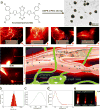
Figure 1
Figure 1. Schematic illustration of biomedical applications using CQS1000 and characterization of CQS1000
a. Schematic illustration of the biological application using CQS1000. b. DLS of CQS1000. c and d. Absorbance and fluorescent emission of CQS1000, demonstrating an absorbance peak at 830 nm and an emission peak at 1,000 nm. The fluorescent emission spectrum was obtained with an 808 nm excitation laser. e. NIR-II signals of CQS1000 with sequential long-pass filters (1,000–1,200 nm).
Figure 2
Figure 2. In vivo fluorescence images of CQS1000(NIR-II) and ICG (NIR-I)
(a)–(c) ICG and CQS1000 were injected intravenously into nude mice and C57BL/6 mice, and then NIR images were obtained at 10 minutes after injection, with the vessel FWHM width (white lines in a, b, c) and SBR (white lines in a, b, c) analysis shown in (c) (failed to acquire because of the broadened peak of signal), (d) and (e), respectively, basing on the cross-sectional intensity profiles. Scale bar: 1.5 cm.
Figure 3
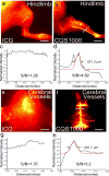
Figure 3. Fluorescence images of the vasculature of hind limb and brain in C57BL/6 mice using CQS1000(NIR-II) and ICG (NIR-I)
In vivo fluorescence vascular image showing hind limb(femoral artery) and cerebrovascular vessels in the NIR-I (ICG) and NIR-II (CQS1000) regions in C57BL/6 mice shown in (a, b, e, f), with the vessel FWHM width and SBR (white lines in a, b, e, f) analysis shown in (c, g) (failed to acquire because of the broadened peak of signal), (d) and (h), respectively, basing on the cross-sectional intensity profiles. Scale bar: 1.5 cm.
Figure 4
Figure 4. In vivo dynamic assessment of angiogenesis development of tumor
NIR-II fluorescence images were captured on the 143B osteosarcoma tumor-bearing mouse every 5 minutes after the CQS1000 injection intravenously (a)–(h). (i). A series analysis of cross-sectional intensity profile measured along the white-dashed line in (a)–(h). (J). A fluorescent intensity analysis representing the intersection point with white-dashed line and the major artery supporting the tumor in (a)–(h), and the half - life of blood circulation was calculated as 88.76 min. (k) and (l). Fluorescent images captured on 14 and 21 days after the 143B osteosarcoma inoculation on nude mice, respectively, indicating the disorganized and tortuous pattern of blood vessel in tumor. Scale bar: 4mm.
Figure 5
Figure 5. Intra-operative assessment of the vascular supply using NIR-II CQS1000 for precise tumor resection
(a)–(d). Pre-evaluation of the blood supply of the tumor. White arrowheads indicate the major blood vessels to supply the tumor. (e) and (f) Intra-operative fluorescent images, indicating that the sentinel lymph node(iliac lymph node) and the concomitant artery were easily exposed and identified (white arrows), respectively. White arrowheads indicate the major blood vessels to supply the tumor. (g) and (h). Major blood vessels supporting the tumor were dissected and exposed (white arrowheads). Major blood vessels were further dissected surgically into two small branches (i) and magnified as (j). As the ex vivo image shown in (k), the major blood artery and vein of tumor were successfully ligated (white arrowheads). Scale bar: 4mm.
Figure 6
Figure 6. In vivo NIR-II images for arterial thrombus formation and incomplete hind limb ischemia
NIR-II images of the left hind limb were captured at 0s (a), 60s (b), 120s (c), 180s (d) after CQS1000 injection intravenously into the tail vein of C57BL/6 mice. White arrows indicate the thrombus formation in femoral artery. NIR-II images of the left hind limb were captured at 0s (e), 60s (f), 120s (g), 180s (h) after CQS1000 injection intravenously into the tail vein of C57BL/6 mice. White arrows indicate the incomplete ischemia (occlusion) in femoral artery. Scale bar: 4mm.
Figure 7
Figure 7. In vivo NIR-II imaging for lymphatic drainage
The popliteal lymph nodes (white arrowhead) could be identified at 1 minute post-injection (a). Then, the inguinal and axillary lymph nodes (white arrowheads) were sequentially and clearly observed during the next 55 minutes after CQS1000 injection (b)–(l). The lymphatic vessels connecting the injection site with the sentinel lymph node (white arrows) as well as the afferent and efferent lymphatic vessels (blue arrowheads) were also unambiguously distinguished. ICG and CQS1000 (40µL) were injected in the left rear footpad of a mouse, respectively. As shown in (m) and (o), the lymph nodes and vessel anatomy identified with CQS1000 are much sharper in comparison with those of ICG, with the FWHM width and SBR (white lines in m, o) analysis shown in (n), and (p), respectively, basing on the cross-sectional intensity profiles. Scale bar: 4mm.
Figure 8
Figure 8. In vivo NIR-II imaging-guided sentinel lymph node mapping and biopsy
The axillary lymph node(white arrow) was clearly identified at 10mins after CQS1000 injection (a). Then, the lymphatic flow toward the sentinel lymph node from the injection site was traced successfully in real time, and then the lymph node (white arrows) was dissected at its precise position in a short time (b)–(i), mimicking the standard SLNB procedure of in clinical cancer surgery. The axillary artery (white arrowhead) was clearly luminous in (e). After the lymph node was resected, the operative field was clearly identified with NIR-II imaging, proving no lymph node was remained except the axillary artery (white arrowhead) (g). SBR was determined with different time after CQS1000 injection (h) and the dissected lymph node was further confirmed by histological analysis (i). Scale bar: 4mm.

Similar articles

Cited by

References

    1. Zhu Q, DeFusco PA, Ricci A, Jr, Cronin EB, Hegde PU, Kane M, Tavakoli B, Xu Y, Hart J, Tannenbaum SH. Radiology. 2013;266:433. - PMC - PubMed
    2. Golijanin J, Amin A, Moshnikova A, Brito JM, Tran TY, Adochite R-C, Andreev GO, Crawford T, Engelman DM, Andreev OA. Proceedings of the National Academy of Sciences. 2016;113:11829. - PMC - PubMed
    3. Sun Y, Ma X, Cheng K, Wu B, Duan J, Chen H, Bu L, Zhang R, Hu X, Deng Z. Angewandte Chemie International Edition. 2015;54:5981. - PubMed
    1. Okochi O, Kaneko T, Sugimoto H, Inoue S, Takeda S, Nakao A. Journal of Surgical Research. 2002;103:109. - PubMed
    1. Meier R, Thuermel K, Noël PB, Moog P, Sievert M, Ahari C, Nasirudin RA, Golovko D, Haller B, Ganter C. Radiology. 2014;270:176. - PubMed
    1. Liu X, Braun GB, Zhong H, Hall DJ, Han W, Qin M, Zhao C, Wang M, She ZG, Cao C. Advanced functional materials. 2016;26:267. - PMC - PubMed
    1. Bu L, Shen B, Cheng Z. Advanced drug delivery reviews. 2014;76:21. - PMC - PubMed
    2. Su X, Cheng K, Wang C, Xing L, Wu H, Cheng Z. Wiley Interdisciplinary Reviews: Nanomedicine and Nanobiotechnology. 2013;5:219. - PubMed

LinkOut - more resources