. 2018 Mar 22:9:180.
doi: 10.3389/fneur.2018.00180. eCollection 2018.

In Situ Proximity Ligation Assay Reveals Co-Localization of Alpha-Synuclein and SNARE Proteins in Murine Primary Neurons

Affiliations

In Situ Proximity Ligation Assay Reveals Co-Localization of Alpha-Synuclein and SNARE Proteins in Murine Primary Neurons

Leire Almandoz-Gil et al. Front Neurol. .

Abstract

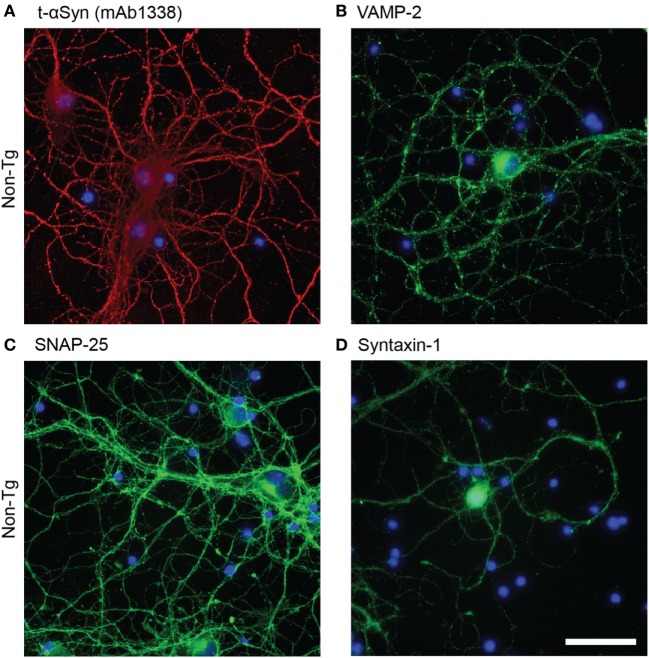
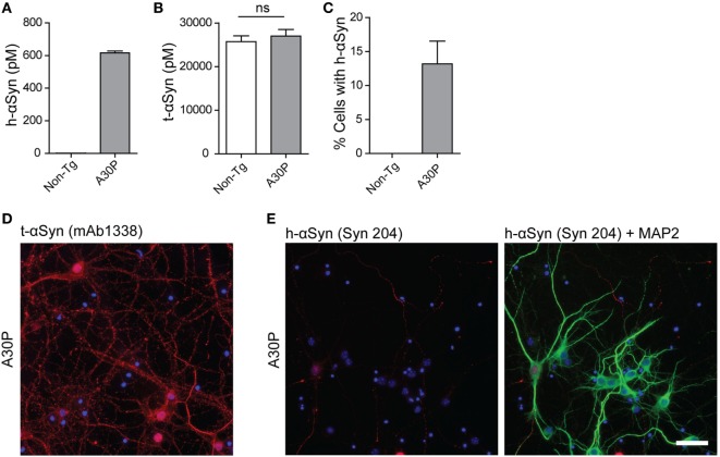
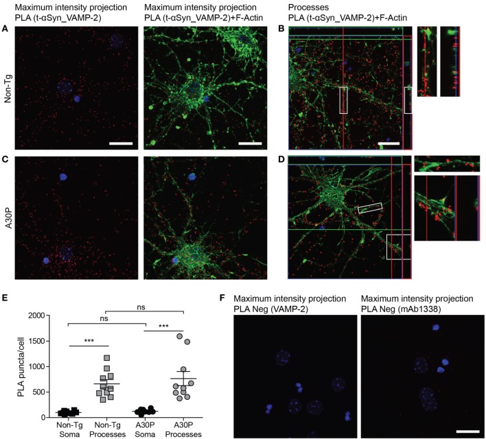
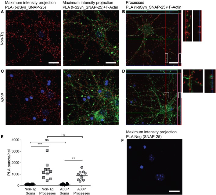
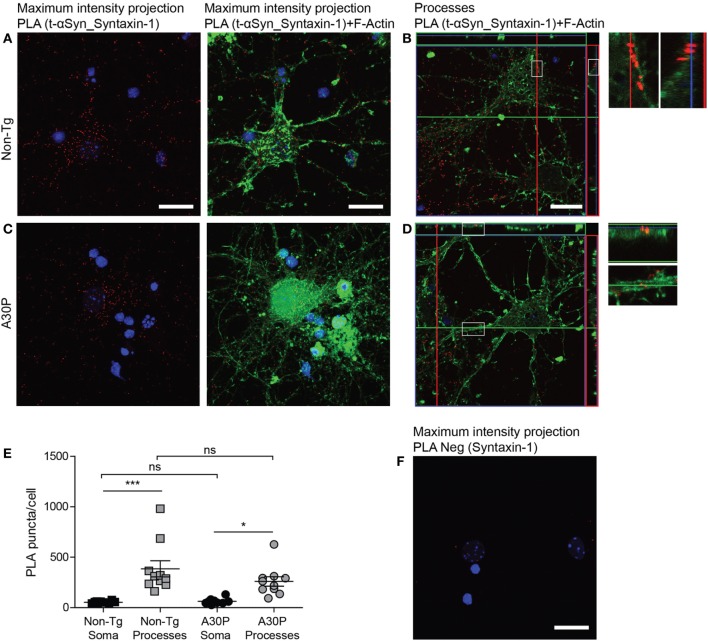
The aggregation of alpha-synuclein (αSyn) is the pathological hallmark of Parkinson's disease, dementia with Lewy bodies and related neurological disorders. However, the physiological function of the protein and how this function relates to its pathological effects remain poorly understood. One of the proposed roles of αSyn is to promote the soluble N-ethylmaleimide-sensitive factor attachment protein receptor (SNARE) complex assembly by binding to VAMP-2. The objective of this study was to visualize the co-localization between αSyn and the SNARE proteins (VAMP-2, SNAP-25, and syntaxin-1) for the first time using in situ proximity ligation assay (PLA). Cortical primary neurons were cultured from either non-transgenic or transgenic mice expressing human αSyn with the A30P mutation under the Thy-1 promoter. With an antibody recognizing both mouse and human αSyn, a PLA signal indicating close proximity between αSyn and the three SNARE proteins was observed both in the soma and throughout the processes. No differences in the extent of PLA signals were seen between non-transgenic and transgenic neurons. With an antibody specific against human αSyn, the PLA signal was mostly located to the soma and was only present in a few cells. Taken together, in situ PLA is a method that can be used to investigate the co-localization of αSyn and the SNARE proteins in primary neuronal cultures.

Keywords: SNAP-25; SNARE; VAMP-2; alpha-synuclein; primary neurons; proximity ligation assay; syntaxin-1.


Figures

Figure 1
Figure 1
Representative images of immunofluorescence staining of non-tg cortical primary neurons (12 DIV) using antibodies against alpha-synuclein (αSyn) [mAb1338, red, (A)], VAMP-2 [(green), (B)], SNAP-25 [(green), (C)], and syntaxin-1 [(green), (D)]. DAPI in blue. Scale bar 50 µm.
Figure 2
Figure 2
Characterization of primary neurons from tg A30P mice. Levels of h-alpha-synuclein (αSyn) in lysates of non-tg and A30P neuronal cultures were measured by ELISA, using the h-αSyn specific antibody 4B12 as a capture antibody (A). Levels of total (t)-αSyn (m-αSyn + h-αSyn) in lysates of non-tg and A30P neuronal cultures were measured by ELISA, using clone 42 as a capture antibody. Two-tailed Student’s t-test, non-significant (ns), error bars represent the SEM (B). Quantification of h-αSyn positive non-tg and A30P neurons, observed by immunofluorescence with h-αSyn specific antibody Syn 204 (C). Representative images of immunofluorescence of tg A30P neurons with t-αSyn mAb1338 (D). Representative images of immunofluorescence with h-αSyn specific antibody Syn 204 of A30P cortical neurons (red). MAP2 neuronal marker (green) and DAPI (blue) (E). Scale bar 50 µm.
Figure 3
Figure 3
Representative confocal images of in situ proximity ligation assay (PLA) between alpha-synuclein (αSyn) (mAb1338) and VAMP-2 (red) in non-tg (A,B) and A30P (C,D) cortical primary neurons. Maximum intensity projections of a confocal z-stack including a whole cell were performed to observe the maximum amount of PLA puncta (A,C). Cross section of the processes allows visualization of the PLA puncta in an orthogonal view [(B,D), inlet], scale bar 20 µm. F-actin stained by phalloidin-FITC (green) and DAPI (blue). Quantification of amount of PLA puncta per cell in the soma and processes. One-way ANOVA followed by Bonferroni post hoc test, non-significant (ns), ***P < 0.001. Error bars represent the SEM (E). Negative control PLA with VAMP-2 antibody only and αSyn antibody (mAb1338) only (F). Scale bar 20 µm.
Figure 4
Figure 4
Representative confocal images of in situ proximity ligation assay (PLA) between alpha-synuclein (αSyn) (mAb1338) and SNAP-25 (red) in non-tg (A,B) and A30P (C,D) cortical primary neurons. Maximum intensity projections of a confocal z-stack including a whole cell were performed to observe the maximum amount of PLA puncta (A,C). Cross section of the processes allows visualization of the PLA puncta in an orthogonal view [(B,D), inlet], scale bar 20 µm. F-actin stained by phalloidin-FITC (green) and DAPI (blue). Quantification of amount of PLA puncta per cell in the soma and processes. One-way ANOVA followed by Bonferroni post hoc test, non-significant (ns), ***P < 0.001. Error bars represent the SEM (E). Negative control PLA with SNAP-25 antibody only (F). Scale bar 20 µm.
Figure 5
Figure 5
Representative confocal images of in situ proximity ligation assay (PLA) between alpha-synuclein (αSyn) (mAb1338) and syntaxin-1 (red) in non-tg (A,B) and A30P (C,D) cortical primary neurons. Maximum intensity projections of a confocal z-stack including a whole cell were performed to observe the maximum amount of PLA puncta (A,C). Cross section of the processes allows visualization of the PLA puncta in an orthogonal view (B,D, inlet), scale bar 20 µm. F-actin stained by phalloidin-FITC (green) and DAPI (blue). Quantification of amount of PLA puncta per cell in the soma and processes. One-way ANOVA followed by Bonferroni post hoc test, non-significant (ns), ***P < 0.001. Error bars represent the SEM (E) Negative control PLA with syntaxin-1 antibody only (F). Scale bar 50 µm.
Figure 6
Figure 6
Representative confocal images of in situ proximity ligation assay (PLA) (red) between h-alpha-synuclein (αSyn) (Syn 204) and VAMP-2 (A,B), h-αSyn (Syn 204) and SNAP-25 (C,D), and h-αSyn (Syn 204) and syntaxin-1 (E,F) in tg A30P neurons and in non-tg neurons (H). Maximum intensity projections of a confocal z-stack including a whole cell were performed to observe the maximum amount of PLA puncta (A,C,E). Cross section of the processes allows visualization of the PLA puncta in an orthogonal view [(B,D,F), inlet], scale bar 20 µm. F-actin stained by phalloidin-FITC (green) and DAPI (blue). Quantification of PLA between h-αSyn and VAMP-2, h-αSyn and SNAP-25, and h-αSyn and syntaxin-1 in A30P neurons (G).
Figure 7
Figure 7
Representative images of NUP98 immunofluorescence staining (green) in non-tg and A30P alpha-synuclein (αSyn) tg primary neurons, DAPI (blue) (A). Representative confocal images (maximum intensity projection) of in situ proximity ligation assay (PLA) in non-tg and A30P cortical primary neurons between αSyn (mAb1338) and the NUP98 (red), DAPI (blue) (B). Scale bar 20 µm.

References

    1. Uversky VN, Li J, Fink AL. Evidence for a partially folded intermediate in alpha-synuclein fibril formation. J Biol Chem (2001) 276:10737–44.10.1074/jbc.M010907200 - DOI - PubMed
    1. Spillantini MG, Schmidt ML, Lee VMY, Trojanowski JQ, Jakes R, Goedert M. Alpha-synuclein in Lewy bodies. Nature (1997) 388:839–40.10.1038/42166 - DOI - PubMed
    1. Gómez-Tortosa E, Newell K, Irizarry MC, Albert M, Growdon JH, Hyman BT. Clinical and quantitative pathologic correlates of dementia with Lewy bodies. Neurology (1999) 53:1284–91.10.1212/WNL.53.6.1284 - DOI - PubMed
    1. Jellinger KA. Neuropathology of sporadic Parkinson’s disease: evaluation and changes of concepts. Mov Disord (2012) 27:8–30.10.1002/mds.23795 - DOI - PubMed
    1. Kramer ML, Schulz-Schaeffer WJ. Presynaptic alpha-synuclein aggregates, not Lewy bodies, cause neurodegeneration in dementia with Lewy bodies. J Neurosci (2007) 27:1405–10.10.1523/JNEUROSCI.4564-06.2007 - DOI - PMC - PubMed

LinkOut - more resources