Review
. 2018 Mar 12:14:603-617.
doi: 10.3762/bjoc.14.47. eCollection 2018.

Enzyme-free genetic copying of DNA and RNA sequences

Affiliations
Review

Enzyme-free genetic copying of DNA and RNA sequences

Marilyne Sosson et al. Beilstein J Org Chem. .

Abstract

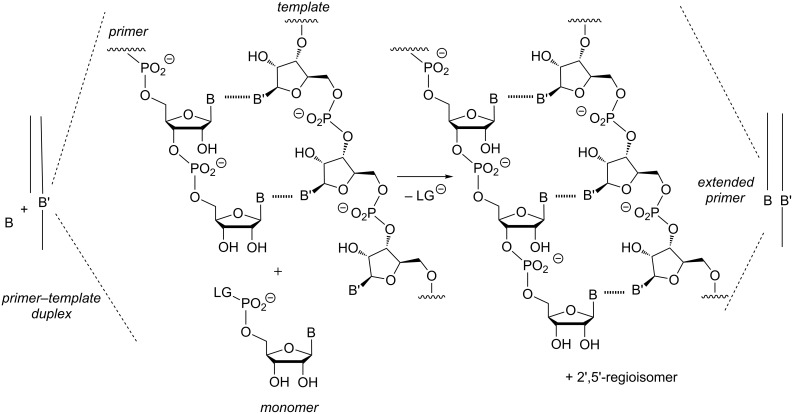
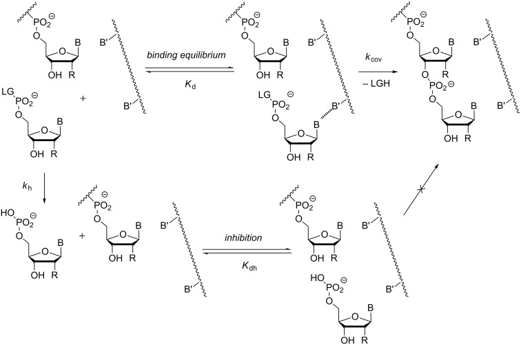
The copying of short DNA or RNA sequences in the absence of enzymes is a fascinating reaction that has been studied in the context of prebiotic chemistry. It involves the incorporation of nucleotides at the terminus of a primer and is directed by base pairing. The reaction occurs in aqueous medium and leads to phosphodiester formation after attack of a nucleophilic group of the primer. Two aspects of this reaction will be discussed in this review. One is the activation of the phosphate that drives what is otherwise an endergonic reaction. The other is the improved mechanistic understanding of enzyme-free primer extension that has led to a quantitative kinetic model predicting the yield of the reaction over the time course of an assay. For a successful modeling of the reaction, the strength of the template effect, the inhibitory effect of spent monomers, and the rate constants of the chemical steps have to be determined experimentally. While challenges remain for the high fidelity copying of long stretches of DNA or RNA, the available data suggest that enzyme-free primer extension is a more powerful reaction than previously thought.

Keywords: DNA; RNA; base pairing; enzyme-free primer extension; nucleotides; oligonucleotides; replication.


Figures

Figure 1
Figure 1
Enzyme-free template-directed extension of an RNA primer by one nucleotide. B = nucleobase, LG = leaving group.
Figure 2
Figure 2
Oligomerization of the 2-methylimidazolide of guanosine-5'-monophosphate on a poly(C) template.
Figure 3
Figure 3
Structures of backbone linkages produced in enzyme-free primer extension reactions: the phosphoramidate of a 3'-amino-2',3'-dideoxynucleoside (1), the phosphoramidate of a 3'-amino-3'-deoxynucleoside (2), the 3',5'-phosphodiester of a natural ribonucleoside (3), and the isomeric 2',5'-phosphodiester of a ribonucleoside (4).
Figure 4
Figure 4
System used for studying the template effect with all 64 possible triplets at the extension site (B/B' = nucleobase).
Figure 5
Figure 5
Interactions attracting the incoming nucleotide to the extension site. Besides base pairing via hydrogen bonding to the templating base, stacking interactions with neighboring bases of primer and a possible downstream-binding helper oligonucleotide, as well as help of solvophobic effects, influence the strength of the template effect.
Figure 6
Figure 6
Three possible fates of activated nucleotides in aqueous buffer that result from hydrolysis, primer extension, and reaction with a free nucleotide, respectively. Other possible pathways, such as cyclization to the 3',5'-cyclic diester or oligomerization are not shown graphically; R is OH for ribonucleotides and is H for deoxynucleotides.
Figure 7
Figure 7
Steps and equilibria considered in our quantitative model of chemical primer extension [34]. The model considers the binding of the activated monomer with its leaving group (LG) to the primer–template complex in the form of the dissociation constant (Kd). It takes into account the rate of hydrolysis with the corresponding rate constant (kh), the binding equilibrium for the hydrolyzed monomer that acts as inhibitor (Kdh), and it assumes a single rate-limiting chemical step (kcov); B, B' = nucleobase = OH for RNA.
Figure 8
Figure 8
Binding equilibrium between mononucleotides and hairpins representing primer–template duplexes, as chosen for measuring dissociation constants by NMR titration.
Figure 9
Figure 9
Template-directed primer extension on an RNA template performed with OAt-GMP at 1.8 mM (orange), 3.6 mM (blue), or 7.2 mM (green) initial concentration. a) Conversion over time, as simulated with our quantitative model, using the dissociation constants of both activated and free nucleotide, and rate constants for hydrolysis and chemical step. The broken black line is the hypothetical conversion of the primer without hydrolysis of monomer and the resulting inhibition; b) Corresponding experimental data, acquired in primer extension assays at 20 °C in buffer (200 mM HEPES, 400 mM NaCl, 80 mM MgCl2, at pH 8.9) at 36 µM primer–template (5'-UAUGCUGG-3' – 3'-CACCCACCACAUACGACCCAAGCACAC-5'); see reference [34] for further details.
Figure 10
Figure 10
Copying of four nucleotides on an immobilized RNA duplex, as reported by Deck et al. [32].
Figure 11
Figure 11
Extension cycle of aminoterminal primer with N-protected nucleotides on solid support, as described by Kaiser et al. [47].
Figure 12
Figure 12
Formation of a highly reactive methylimidazolium bisphosphate from methylimidazolides of nucleotides.
Figure 13
Figure 13
31P NMR spectrum (161.9 MHz) of crude MeIm-GMP in D2O. The resonance of the imidazolium bisphosphate appears at −10.8 ppm, and that of the pyrophosphate GppG at −11.3 ppm.
Figure 14
Figure 14
Imidazolium bisphosphate as intermediate in the primer extension reaction, as described by Szostak and colleagues. a) Intermediate of an extension with aminoimidazolides as monomers [52]; b) one of the structural arrangements found in a recent crystallography study that used oligophosphates as model compounds [53].
Figure 15
Figure 15
Proposed steps of enzyme-free primer extension with in situ activation, using the "general condensation buffer" containing EDC and 1-ethylimidazole as organocatalyst.

References

    1. Kornberg A, Baker T A. DNA Replication. 2nd ed. Mill Valley: University Science Books; 2005.
    1. Crick F. Nature. 1970;227:561–563. doi: 10.1038/227561a0. - DOI - PubMed
    1. Cech T R. Proc Natl Acad Sci U S A. 1986;83:4360–4363. doi: 10.1073/pnas.83.12.4360. - DOI - PMC - PubMed
    1. Kruger K, Grabowski P J, Zaug A J, Sands J, Gottschling D E, Cech T R. Cell. 1982;31:147–157. doi: 10.1016/0092-8674(82)90414-7. - DOI - PubMed
    1. Wochner A, Attwater J, Coulson A, Holliger P. Science. 2011;332:209–212. doi: 10.1126/science.1200752. - DOI - PubMed

LinkOut - more resources