. 2017 Nov 20;4(11):1337-1343.
doi: 10.1364/OPTICA.4.001337.

Focusing light inside scattering media with magnetic-particle-guided wavefront shaping

Affiliations

Focusing light inside scattering media with magnetic-particle-guided wavefront shaping

Haowen Ruan et al. Optica. .

Abstract

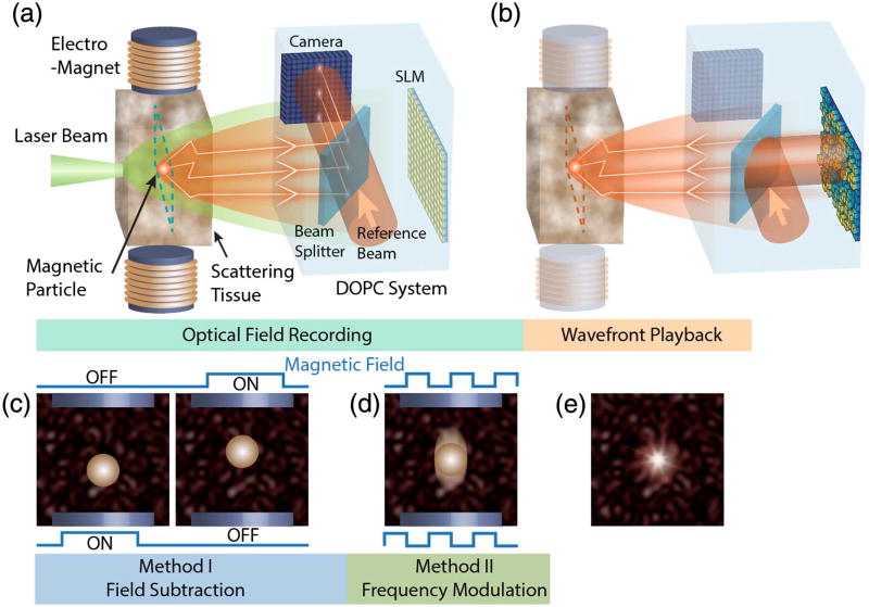
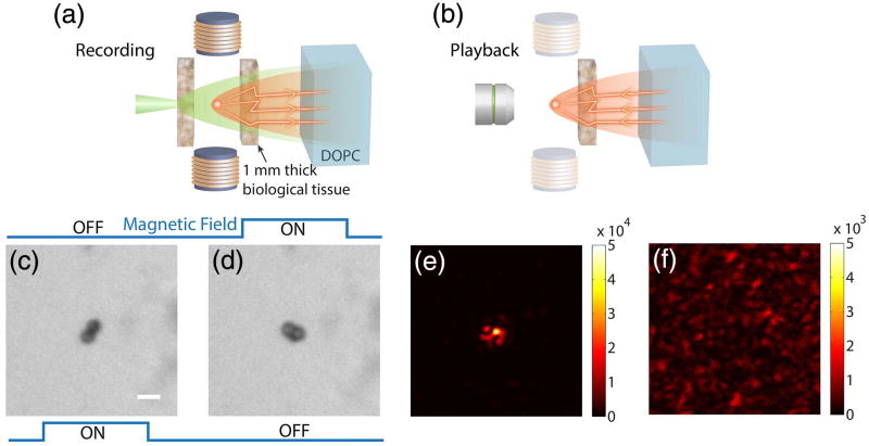
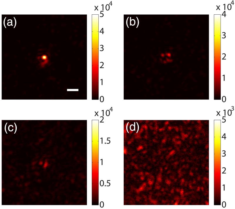
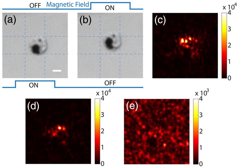
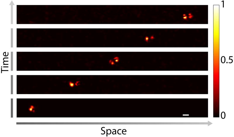
Optical scattering has traditionally limited the ability to focus light inside scattering media such as biological tissue. Recently developed wavefront shaping techniques promise to overcome this limit by tailoring an optical wavefront to constructively interfere at a target location deep inside scattering media. To find such a wavefront solution, a "guide-star" mechanism is required to identify the target location. However, developing guidestars of practical usefulness is challenging, especially in biological tissue, which hinders the translation of wavefront shaping techniques. Here, we demonstrate a guidestar mechanism that relies on magnetic modulation of small particles. This guidestar method features an optical modulation efficiency of 29% and enables micrometer-scale focusing inside biological tissue with a peak intensity-to-background ratio (PBR) of 140; both numbers are one order of magnitude higher than those achieved with the ultrasound guidestar, a popular guidestar method. We also demonstrate that light can be focused on cells labeled with magnetic particles, and to different target locations by magnetically controlling the position of a particle. Since magnetic fields have a large penetration depth even through bone structures like the skull, this optical focusing method holds great promise for deep-tissue applications such as optogenetic modulation of neurons, targeted light-based therapy, and imaging.


Figures

Fig. 1
Fig. 1
Principle of magnetic-particle-guided optical focusing. (a) A magnetic particle is embedded in a piece of scattering tissue. A portion of the impinging laser beam interacts with the particle and the resulting tagged light is detected interferometrically using the camera of a DOPC system. (b) After capturing the field of the tagged light, the conjugate wavefront is displayed on the spatial light modulator (SLM) of the DOPC system. The reconstructed conjugate light field then retraces the scattering paths and forms a focus at the location of the magnetic particle. Panels (c) and (d) show two methods to separate the tagged light field from the background unmodulated light. The field subtraction method in (c) captures two optical fields before and after a magnetic field displaces the magnetic particle. The differential field nullifies the contribution from the background, which is not scattered by the particle. The frequency modulation method shown in (d) uses an AC magnetic field to make the magnetic particle oscillate, which shifts the frequency of the light, which interacts with the particle. By matching the frequency of a planar reference beam with that of the tagged light, the DOPC system detects the tagged light field via phase-shifting holography. (e) After imprinting the conjugate wavefront of the tagged light on a planar reference beam using the SLM, the conjugate wave forms a bright focus on top of a dim background at the location of the magnetic particle inside the scattering medium.
Fig. 2
Fig. 2
Magnetic-particle-guided optical focusing with the field subtraction method. (a) Schematic of the setup to record the field of the tagged light. (b) Schematic of the setup for playback of the tagged field and observation of the focus. In this step, the tissue on the left side was removed and an imaging system was used to observe the light intensity distribution on the magnetic particle plane. Panels (c) and (d) show bright-field images of the particles with the magnetic field in different directions. (e) The focus observed with the setup shown in (b). (f) Control experiment: no focus was observed when the magnetic fields were turned off and the experiment was repeated. Scale bar, 5 µm.
Fig. 3
Fig. 3
Magnetic-particle-guided optical focusing with the frequency modulation method. The electromagnets were driven by 25 Hz rectangular waves. Images were captured with the setup shown in Fig. 2(b). The focus achieved when the reference beam frequency was shifted by (a) 25 Hz (fundamental frequency), (b) 50 Hz (second harmonic), and (c) 75 Hz (third harmonic) relative to the laser frequency. (d) Control experiment: no focus was observed when the reference beam frequency was shifted by 30 Hz (frequency mismatch). Scale bar, 5 µm.
Fig. 4
Fig. 4
Focusing light onto a targeted cell that endocytosed magnetic particles of 453 nm diameter. Panels (a) and (b) show bright-field images of a cell under two magnetic fields. (c) Focus achieved by the field subtraction method. (d) Focus achieved by the frequency modulation method (fm = 25 Hz). (e) Control experiment: no focus was observed when the SLM pattern was circularly shifted by 10 × 10 pixels after obtaining the result in (d). Scale bar, 5 µm.
Fig. 5
Fig. 5
Focusing light to different target locations by controlling the positions of the magnetic particles using an external magnetic field. The magnetic particles were driven to the target locations inside a microfluidic channel based on the position feedback from the observation microscope [Fig. 2(b)]. After reaching each target location, the magnetic particles were covered by the scattering samples on both sides as shown in Fig. 2(a), and the DOPC process was performed to create a focus through the scattering sample on the DOPC system side. Then, the scattering sample on the observation microscope side was removed [Fig. 2(b)], and the focus was observed directly. Scale bar, 5 µm.

References

    1. Ntziachristos V. Going deeper than microscopy: the optical imaging frontier in biology. Nat. Methods. 2010;7:603–614. - PubMed
    1. Vellekoop IM, Mosk AP. Focusing coherent light through opaque strongly scattering media. Opt. Lett. 2007;32:2309–2311. - PubMed
    1. Vellekoop IM. Feedback-based wavefront shaping. Opt. Express. 2015;23:12189–12206. - PubMed
    1. Mosk AP, Lagendijk A, Lerosey G, Fink M. Controlling waves in space and time for imaging and focusing in complex media. Nat. Photonics. 2012;6:283–292.
    1. Horstmeyer R, Ruan H, Yang C. Guidestar-assisted wavefront-shaping methods for focusing light into biological tissue. Nat. Photonics. 2015;9:563–571. - PMC - PubMed

LinkOut - more resources