. 2018 Jul;223(6):2999-3006.
doi: 10.1007/s00429-018-1661-x. Epub 2018 Apr 6.

Frontal cortical control of posterior sensory and association cortices through the claustrum

Affiliations

Frontal cortical control of posterior sensory and association cortices through the claustrum

Michael G White et al. Brain Struct Funct. 2018 Jul.

Abstract

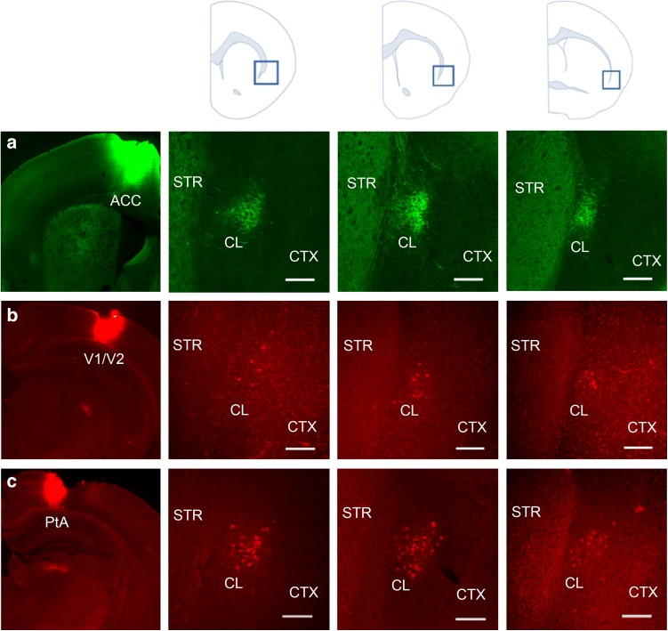
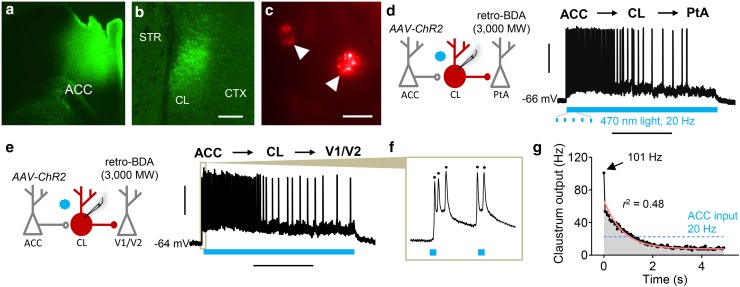
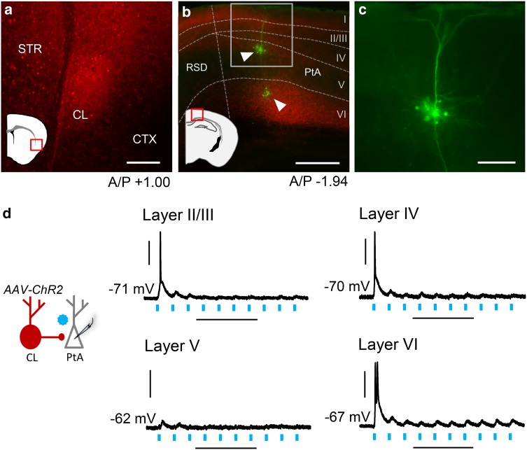
The claustrum is a telencephalic gray matter nucleus that is richly interconnected with the neocortex. This structure subserves top-down executive functions that require frontal cortical control of posterior cortical regions. However, functional anatomical support for the claustrum allowing for long-range intercortical communication is lacking. To test this, we performed a channelrhodopsin-assisted long-circuit mapping strategy in mouse brain slices. We find that anterior cingulate cortex input to the claustrum is transiently amplified by claustrum neurons that, in turn, project to parietal association cortex or to primary and secondary visual cortices. Additionally, we observe that claustrum drive of cortical neurons in parietal association cortex is layer-specific, eliciting action potential generation briefly in layers II/III, IV, and VI but not V. These data are the first to provide a functional anatomical substrate through claustrum that may underlie top-down functions, such as executive attention or working memory, providing critical insight to this most interconnected and enigmatic nucleus.

Keywords: Anterior cingulate cortex; Macrocircuit; Optogenetics; Parietal association cortex; Top-down; Visual cortices.


Conflict of interest statement

Conflict of interest

The authors declare no financial and non-financial competing interests.

Ethical standards

All applicable international, national, and/or institutional guidelines for the care and use of animals were followed. All procedures performed in studies involving animals were in accordance with the ethical standards of the institution or practice at which the studies were conducted.

Figures

Fig. 1
Fig. 1
Claustrum receives dense innervation from anterior cingulate cortex (ACC) and projects to visual cortices (V1/V2) and parietal association cortex (PtA). a Left: photomicrograph showing anterograde neuronal tract tracer biotinylated dextran amine (BDA, 10,000 MW; green) injected into ACC. Right: BDA-labeled terminals (green) from ACC densely innervated contralateral claustrum at all three rostrocaudal levels of claustrum as depicted by the blue boxes in the anatomical cartoons above. b Left: photomicrograph showing retrograde tract tracer cholera toxin B subunit (CTb; red) injected into V1/V2. Right: CTb-labeled cells projecting to V1/V2 were found at all three rostrocaudal levels of ipsilateral claustrum. c Left: photomicrograph showing CTb (red) injected into PtA. Right: a dense population of CTb-labeled cells projecting to PtA were found at all three rostrocaudal levels of ipsilateral claustrum. STR striatum, CL claustrum, CTX cortex. Scale bars—200 µm
Fig. 2
Fig. 2
Claustrum neurons projecting to PtA and V1/V2 receive and transiently amplify ACC input. a Photomicrograph showing adeno-associated virus expressing channelrhodopsin-2 (AAV-ChR2) with eYFP tag injected into ACC. b ACC afferents expressing ChR2 (green) densely labeled the claustrum. c PtA projection neurons in claustrum (arrows) were labeled with retrogradely trafficked BDA (retro-BDA; 3000 MW) that was injected into PtA. d Left: ChR2-assisted macrocircuit mapping strategy entailed optogenetic stimulation of ACC afferents (AAV-ChR2) with 470 nm light (blue star) while performing whole-cell recordings from claustrum (CL) projection neurons to PtA. Right: representative trace showing a claustrum neuron projecting to PtA that initially burst-fired in response to a 5 s train of 20 Hz optogenetic ACC stimulation. Firing diminished as the stimulation train progressed. e The same macrocircuit mapping strategy as in d was employed to target claustrum neurons projecting to V1/V2 while recording responses to ACC afferent stimulation. Right: representative trace showing a claustrum neuron projecting to V1/V2 that initially burst-fired in response to a 5 s train of 20 Hz ACC stimulation. Firing diminished over time. f Representative trace showing a V1/V2-projecting neuron firing multiple action potentials in response to single pulses of ACC afferent stimulation. g Graph showing average output of V1/V2- and PtA-projecting claustrum neurons over time in response to a constant ACC input (20 Hz for 5 s). Peak average output was at the first light pulse (101 Hz) and exponentially decayed over the light train (n = 15; r2 = 0.48, p < 0.005). Horizontal scale bars—200 µm b; 25 µm c; 2 s d, e. Vertical scale bars—30 mV
Fig. 3
Fig. 3
Claustrum afferent stimulation drives PtA neuron firing in layers II/III, IV, and VI but not V. a Photomicrograph showing AAV-ChR2 (red) expression after injection into the claustrum. The photomicrograph location is indicated by the red box on the cartoon. b Photomicrograph showing PtA layer-specific innervation of claustrum afferents expressing ChR2 (red box shows PtA inset region). Neurons across PtA layers were targeted for whole-cell recordings and filled with neurobiotin (green, arrowheads). Cortical layers were delineated by staining with 4′,6-diamidine-2′-phenylindole (DAPI, not shown). Innervation of layer V was relatively sparse compared to other cortical layers. c Inset from b showing PtA layer V pyramidal neuron at high magnification. d Left: schematic showing claustrum innervation of PtA layers. Right: representative traces showing responses to optogenetic stimulation of claustrum afferents (20 Hz, 470 nm light). Action potential firing was noted at initial light pulses across PtA layers except for layer V. Horizontal scale bars—200 µm a; 400 µm b; 100 µm c; 200 ms d. Vertical scale bars—30 mV. A/P anterior/posterior, RSD retrosplenial dysgranular cortex

References

    1. Arcaro MJ, Pinsk MA, Kastner S. The anatomical and functional organization of the human visual pulvinar. J Neurosci. 2015;35:9848–9871. doi: 10.1523/JNEUROSCI.1575-14.2015. - DOI - PMC - PubMed
    1. Atlan G, Terem A, Peretz-Rivlin N, Groysman M, Citri A. Mapping synaptic cortico-claustral connectivity in the mouse. J Comp Neurol. 2017;525:1381–1402. doi: 10.1002/cne.23997. - DOI - PubMed
    1. Barbas H, Rempel-Clower N. Cortical structure predicts the pattern of corticocortical connections. Cereb Cortex. 1997;7:635–646. doi: 10.1093/cercor/7.7.635. - DOI - PubMed
    1. Benevento LA, Rezak M. The cortical projections of the inferior pulvinar and adjacent lateral pulvinar in the rhesus monkey (Macaca mulatta): an autoradiographic study. Brain Res. 1976;108:1–24. doi: 10.1016/0006-8993(76)90160-8. - DOI - PubMed
    1. Braak H, Braak E. Neuronal types in the claustrum of man. Anat Embryol (Berl) 1982;163:447–460. doi: 10.1007/BF00305558. - DOI - PubMed

MeSH terms

LinkOut - more resources