. 2018 Apr 4;3(2):e00038-18.
doi: 10.1128/mSphere.00038-18. Print 2018 Apr 25.

Pseudotyping of HIV-1 with Human T-Lymphotropic Virus 1 (HTLV-1) Envelope Glycoprotein during HIV-1-HTLV-1 Coinfection Facilitates Direct HIV-1 Infection of Female Genital Epithelial Cells: Implications for Sexual Transmission of HIV-1

Affiliations

Pseudotyping of HIV-1 with Human T-Lymphotropic Virus 1 (HTLV-1) Envelope Glycoprotein during HIV-1-HTLV-1 Coinfection Facilitates Direct HIV-1 Infection of Female Genital Epithelial Cells: Implications for Sexual Transmission of HIV-1

Yuyang Tang et al. mSphere. .

Abstract

Female genital epithelial cells cover the genital tract and provide the first line of protection against infection with sexually transmitted pathogenic viruses. These cells normally are impervious to HIV-1. We report that coinfection of cells by HIV-1 and another sexually transmitted virus, human T-lymphotropic virus 1 (HTLV-1), led to production of HIV-1 that had expanded cell tropism and was able to directly infect primary vaginal and cervical epithelial cells. HIV-1 infection of epithelial cells was blocked by neutralizing antibodies against the HTLV-1 envelope (Env) protein, indicating that the infection was mediated through HTLV-1 Env pseudotyping of HIV-1. Active replication of HIV-1 in epithelial cells was demonstrated by inhibition with anti-HIV-1 drugs. We demonstrated that HIV-1 derived from peripheral blood of HIV-1-HTLV-1-coinfected subjects could infect primary epithelial cells in an HTLV-1 Env-dependent manner. HIV-1 from subjects infected with HIV-1 alone was not able to infect epithelial cells. These results indicate that pseudotyping of HIV-1 with HTLV-1 Env can occur in vivo Our data further reveal that active replication of both HTLV-1 and HIV-1 is required for production of pseudotyped HIV-1. Our findings indicate that pseudotyping of HIV-1 with HTLV-1 Env in coinfected cells enabled HIV-1 to directly infect nonpermissive female genital epithelial cells. This phenomenon may represent a risk factor for enhanced sexual transmission of HIV-1 in regions where virus coinfection is common.IMPORTANCE Young women in certain regions of the world are at very high risk of acquiring HIV-1, and there is an urgent need to identify the factors that promote HIV-1 transmission. HIV-1 infection is frequently accompanied by infection with other pathogenic viruses. We demonstrate that coinfection of cells by HIV-1 and HTLV-1 can lead to production of HIV-1 pseudotyped with HTLV-1 Env that is able to directly infect female genital epithelial cells both in vitro and ex vivo Given the function of these epithelial cells as genital mucosal barriers to pathogenic virus transmission, the ability of HIV-1 pseudotyped with HTLV-1 Env to directly infect female genital epithelial cells represents a possible factor for increased risk of sexual transmission of HIV-1. This mechanism could be especially impactful in settings such as Sub-Saharan Africa and South America, where HIV-1 and HTLV-1 are both highly prevalent.

Keywords: envelope glycoprotein; epithelial cells; human T-cell leukemia virus; human immunodeficiency virus; primary T-cells; pseudotype; retroviruses; sexual transmission; virus tropism.


Figures

FIG 1
FIG 1
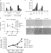
Coreplication of HIV-1 and HTLV-1 in primary CD4+ T cells. Primary CD4+ T lymphocytes were infected with HIV-1 IIIB or Bal and/or HTLV-1 as indicated. (A) HIV-1 IIIB or Bal and HTLV-1 DNA in infected cells at day 4 (HIV-1) or 5 (HTLV-1) postexposure was quantified by qPCR. (B) HTLV-1 production in culture supernatant at days 4, 6, and 8 postinfection was determined by qRT-PCR. (C) HIV-1 release at days 4, 6, and 8 postinfection was quantified by p24 ELISA. **, P < 0.001 (in comparison with data obtained with cells infected with HIV-1 alone). (D) Representative images show that exposure to HTLV-1 resulted in morphological changes in primary CD4+ T lymphocytes. Images were taken at day 5 after HTLV-1 exposure. (E) HTLV-1 exposure stimulated primary CD4+ T lymphocyte proliferation. The data shown represent the mean of data from three to six independent experiments.
FIG 2
FIG 2
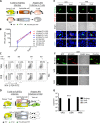
Infection of epithelial cells with HIV-1 from HIV-1–HTLV-1-coinfected CD4+ T cells. TANI reporter cells and primary epithelial cells isolated from VAG and CER biopsy tissues were infected by coculture with HIV-1 (Bal or IIIB)- and HTLV-1-coinfected (HTLV+), HIV-1-monoinfected (HTLV), or mock-infected (HTLV HIV) primary CD4+ T cells. The viruses used to infect donor primary CD4+ T cells are indicated in each panel. (A) HIV-1 infection of TANI reporter cells as indicated by GFP expression (green) and HIV-1 Gag expression (red) after immunostaining with anti-HIV-1 Gag antibody. (B, C) HIV-1 infection of primary CER (B) and VAG (C) epithelial cells. At day 5 postinfection, staining was performed with anti-HIV-1 Gag (green) and anti-CK19 (blue) antibodies, followed by confocal immunofluorescence microscopy analysis. The bright and merged views of the same field are shown (bottom). (D, E) DNA levels of both retroviruses in infected primary CER (D) and VAG (E) epithelial cells at day 5 postexposure were determined by qPCR. The data shown represent the mean ± the standard deviation from three independent experiments. Scale bars, 20 µm.
FIG 3
FIG 3
Infection of female genital epithelial cells by cell-free HIV-1 from HIV-1–HTLV-1-coinfected CD4+ T cells. TANI, CER, and VAG cells were infected with progeny virus from HIV-1 (IIIB or Bal)- and HTLV-1-coinfected primary CD4+ T cells or infected with progeny virus from control CD4+ T cells infected with HIV-1 or HTLV-1 alone or mock infected. Viruses used to infect donor primary CD4+ T cells are indicated in each panel. (A) TANI cells were stained with anti-HIV-1 Gag antibody and analyzed for GFP (green) and HIV-1 Gag (red) expression. Scale bars, 30 µm. (B, C) Primary CER and VAG epithelial cells were stained with anti-HIV-1 p24Gag (green) and anti-CK19 (blue) antibodies. Scale bars, 20 µm. (D) HIV-1 and HTLV-1 release into culture supernatants of CER and VAG primary epithelial cells, as well as TANI cells, was quantified by qRT-PCR at day 5 postinfection. The data shown represent the mean ± the standard deviation of data from three independent experiments.
FIG 4
FIG 4
Infection with HTLV-1-pseudotyped HIV-1 was strongly inhibited by HTLV-1 neutralizing antibodies and HIV-1 inhibitor treatments. (A, B) TANI or primary VAG or CER epithelial cells were cocultured with HIV-1 IIIB–HTLV-1-coinfected T cells in the presence of antibodies as indicated. (A) De novo HTLV-1 DNA from infected TANI, CER, and VAG primary epithelial cells at day 5 postinfection was quantified by qPCR (normalized to the GAPDH internal control). (B) The effect of the antibodies on HIV-1 infection of epithelial cells was determined either by quantifying GFP-positive cells (TANI) or by quantifying the de novo level of HIV-1 DNA normalized to the GAPDH internal control (CER and VAG epithelial cells). PRH4 and PRH7A, neutralizing MAbs against HTLV-1 gp46; PRH1, HTLV-1 gp46 binding antibody without neutralizing activity; HAM serum, IgG from the serum of patients with HAM; 2G12, neutralizing MAb against HIV-1 gp120. Viral replication in mock-treated epithelial cells was set as 1.0. *, P < 0.05; **, P < 0.001. For PRH4 and PRH7A, data were compared to mouse IgG-treated samples. For HAM serum, data were compared to normal human IgG-treated samples. The data represent the mean ± the standard deviation of data from three independent experiments. The concentration used for PHR4, PRH7A, PRH1, and mouse IgG was 10 µg/ml. The concentration used for HAM serum IgG and normal human IgG was 100 µg/ml. The concentration used for 2G12 was 16 µg/ml. (C) HeLa cells and primary CER and VAG epithelial cells and were infected with HIV-1 by coculture with HIV-1 IIIB–HTLV-1-coinfected CD4+ T cells in the presence of AZT (10 µM), SAQ (0.4 µM), or DAR (0.5 µM). HIV-1 release into culture supernatants of epithelial cells was determined by p24 ELISA at days 4 and 5 postinfection. The data represent the mean ± the standard deviation of data from three independent experiments.
FIG 5
FIG 5
Genital epithelial cells infected with HIV-1 from HTLV-1-coinfected T cells efficiently transmit HIV-1 to susceptible T cells and also produce pseudotyped HIV-1. (A) Cartoon illustrating the system used to test the ability of primary epithelial cells HIV-1 infected through pseudotyping to produce infectious HIV-1. (B, C) The progeny virus from epithelial cells infected by coculture with infected T cells was added to naive PM1 cells. The viruses used to infect donor T cells and types of epithelial cells are as indicated. HIV-1 infection of PM1 cells was determined at days 4 and 6 postinfection by measuring HIV-1 release by p24 Gag ELISA (B) and at day 6 postexposure by quantifying HIV-1 p24 Gag-positive cells by flow cytometry (C). (D) Cartoon illustrating the system used to test whether HIV-1–HTLV-1-coinfected donor T cells can transmit both HIV-1 and HTLV-1 to epithelial cells, resulting in subsequent rounds of epithelial cell infection with pseudotyped HIV-1. SAQ and DAR should inhibit pseudotyped HIV-1 infection of epithelial cells and subsequent pseudotyped HIV-1 production. (E) HIV-1–HTLV-1-coinfected T cells transmitted both HIV-1 and HTLV-1 to epithelial cells. CER and VAG epithelial cells were infected by coculture with HIV-1 IIIB–HTLV-1-infected T cells and stained at day 5 postinfection for HIV-1 Gag (green), HTLV-1 p19 (red), and CK19. Mono, HIV-1 monoinfection; dual, HIV-1–HTLV-1 coinfection. Scale bars, 20 µm. (F, G) Epithelial cells were cocultured with HIV-1 IIIB–HTLV-1-coinfected T cells with or without the drugs indicated. Progeny HIV-1 from epithelial cells was added to TANI cells. (F) Images showing HIV-1 infection of TANI cells after exposure to progeny virus from infected CER cells as determined by analyzing GFP (green) and HIV-1 Gag expression (red). Scale bars, 20 µm. (G) HIV-1 release in culture supernatants from infected TANI cells at day 5 postexposure determined by qRT-PCR. The data represent the mean ± the standard deviation from three independent experiments.
FIG 6
FIG 6
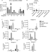
PBMC-associated HIV-1 from HIV-1–HTLV-1-coinfected patients infects epithelial cells. (A) HTLV-1 RNA released into culture supernatants of PBMCs from selected visits of HIV-1-infected, HTLV-1-seropositive subjects following culture for 2 days was determined by HTLV-1 specific qRT-PCR. S, subject; V, visit. (B to D) PBMCs from patients were used to infect epithelial reporter (TANI) cells by coculture. HIV-1 infection of TANI cells was determined by measuring GFP and HIV-1 Gag expression. (B) Representative images show that HIV-1 from PBMCs of HIV-1–HTLV-1-coinfected subject 2 at visit 3 and subject 5 at visits 7 and 24 of HIV-1–HTLV-1-coinfected groups were able to infect TANI reporter cells. Scale bars, 50 µm. (C, D) PBMCs from the indicated person visits of the HIV-1–HTLV-1 dually infected group (C) and from 18 HIV-1-monoinfected control subjects (D) were used to infected TANI cells. The number of HIV-1-infected TANI cells in a 1.5-cm2 area of the glass bottom dishes was quantified.
FIG 7
FIG 7
Reactivation of latent HTLV-1 in PBMCs from HIV-1–HTLV-1-coinfected subjects. (A) HTLV-1 DNA in PBMCs from selected person visits of the HIV-1–HTLV-1-coinfected group was quantified by qPCR. (B) PBMCs from the selected person visits of the HIV-1–HTLV-1-coinfected group were treated with the compounds indicated. HTLV-1 RNA in the culture supernatants was quantified at day 2 posttreatment. The final concentrations of the compounds used were as follows: PEP005, 20 nM; JQ1, 0.5 µM; PHA, 2 µg/ml; PMA 25 ng, PMA at 25 ng/ml plus ionomycin at 5 µM; PMA 12.5 ng, PMA at 12.5 ng/ml plus ionomycin at 5 µM; SAHA, 500 nM; CD3/CD28/CD2, 1× Stem Cell stock. (C) HTLV-1 RNA level in the supernatants of the PBMCs from each selected person visit of HIV-1–HTLV-1-coinfected donors after 2 days of stimulation with the compounds indicated. Data represent the mean ± the standard deviation from triplicate experiments. S, subject; V, visit.
FIG 8
FIG 8
PBMCs from HIV-1-infected patients with latent HTLV-1 infection transmitted HIV-1 to epithelial reporter cells after reactivation of HTLV-1. PBMCs from HIV-1–HTLV-1-coinfected patients were treated with the compounds indicated for 2 days and then cocultured with TANI reporter cells. HIV-1 infection of TANI cells was determined by measuring GFP (green) and HIV-1 Gag (red) expression. (A) Representative images showing HIV-1 infection of TANI cells after coculture with PEP005- and JQ1-treated PBMCs from HIV-1–HTLV-1 dually infected subject 2 at visits 3 and 17. (B) Enhanced HIV-1 infection of TANI cells after stimulation of HTLV-1 replication in PBMCs from a HIV-1–HTLV-1-coinfected individual. TANI cells were cocultured with mock-treated or with PEP005- and JQ1-treated PBMCs from subject 2 at visit 3. The number of HIV-1-infected TANI cells in a 1.5-cm2 area of the glass bottom dish was quantified. HIV-1-positive TANI cells cocultured with mock-treated PBMCs were set as 100. (C, D) Representative images of HIV-1 infection of TANI cells after coculture with PBMCs from HIV-1–HTLV-1-coinfected subject 3 at visit 1 (C) and subject 1 at visit 3 (D) treated with the compound indicated or mock treated. Scale bars, 20 µm.

Similar articles

Cited by

References

    1. Royce RA, Seña A, Cates W Jr., Cohen MS. 1997. Sexual transmission of HIV. N Engl J Med 336:1072–1078. doi:10.1056/NEJM199704103361507. - DOI - PubMed
    1. Bobardt MD, Chatterji U, Selvarajah S, Van der Schueren B, David G, Kahn B, Gallay PA. 2007. Cell-free human immunodeficiency virus type 1 transcytosis through primary genital epithelial cells. J Virol 81:395–405. doi:10.1128/JVI.01303-06. - DOI - PMC - PubMed
    1. Cameron PU, Freudenthal PS, Barker JM, Gezelter S, Inaba K, Steinman RM. 1992. Dendritic cells exposed to human immunodeficiency virus type-1 transmit a vigorous cytopathic infection to CD4+ T cells. Science 257:383–387. doi:10.1126/science.1352913. - DOI - PubMed
    1. Gray RH, Wawer MJ, Brookmeyer R, Sewankambo NK, Serwadda D, Wabwire-Mangen F, Lutalo T, Li X, vanCott T, Quinn TC, Rakai Project Team . 2001. Probability of HIV-1 transmission per coital act in monogamous, heterosexual, HIV-1-discordant couples in Rakai, Uganda. Lancet 357:1149–1153. doi:10.1016/S0140-6736(00)04331-2. - DOI - PubMed
    1. Boily MC, Baggaley RF, Wang L, Masse B, White RG, Hayes RJ, Alary M. 2009. Heterosexual risk of HIV-1 infection per sexual act: systematic review and meta-analysis of observational studies. Lancet Infect Dis 9:118–129. doi:10.1016/S1473-3099(09)70021-0. - DOI - PMC - PubMed

Publication types

MeSH terms

LinkOut - more resources