. 2018 Apr 6;13(4):e0195165.
doi: 10.1371/journal.pone.0195165. eCollection 2018.

Toll-like receptor 3 (TLR3) promotes the resolution of Chlamydia muridarum genital tract infection in congenic C57BL/6N mice

Affiliations

Toll-like receptor 3 (TLR3) promotes the resolution of Chlamydia muridarum genital tract infection in congenic C57BL/6N mice

Sebastian E Carrasco et al. PLoS One. .

Abstract

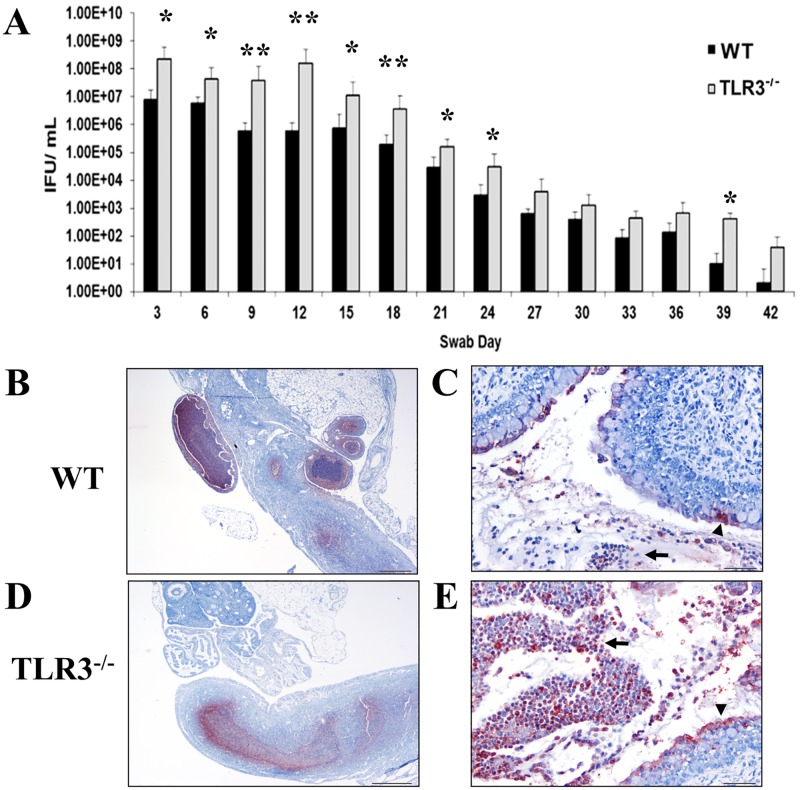
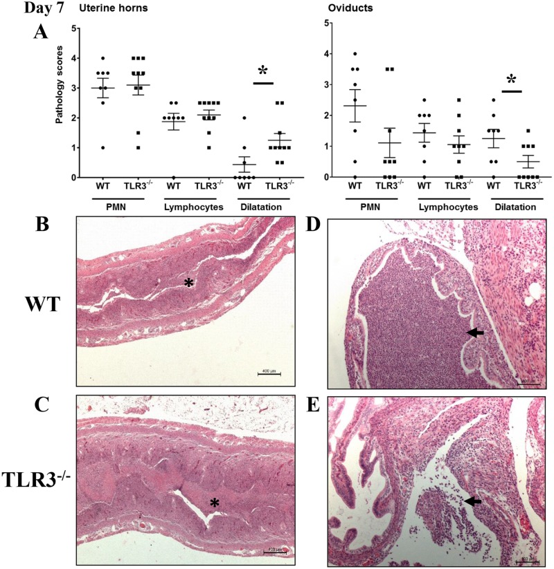
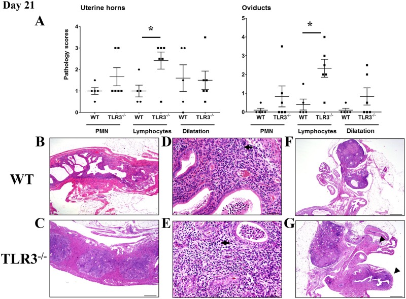
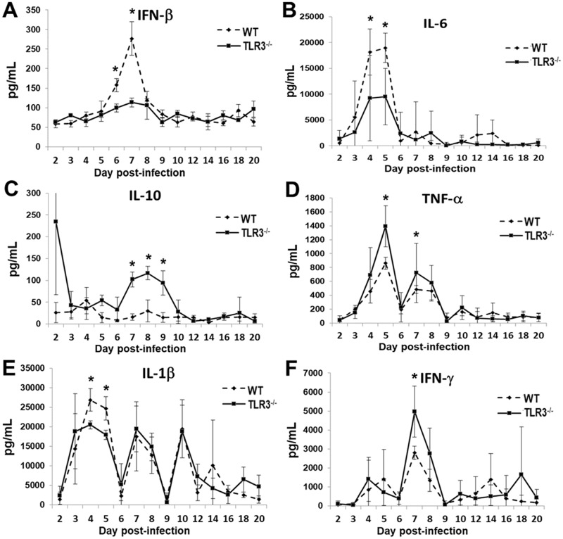
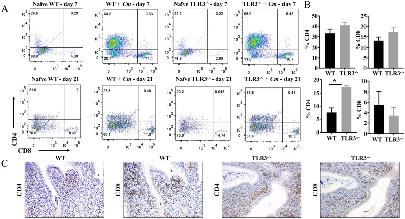
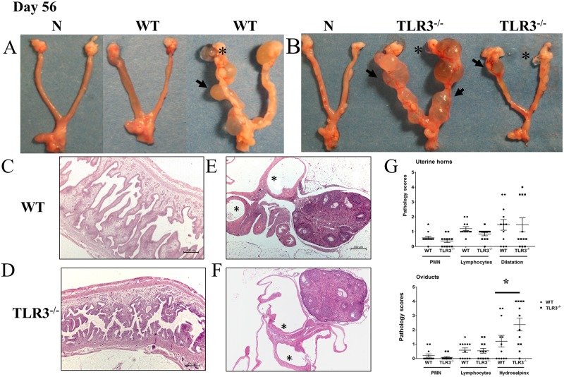
Chlamydia trachomatis urogenital serovars primarily replicate in epithelial cells lining the reproductive tract. Epithelial cells recognize Chlamydia through cell surface and cytosolic receptors, and/or endosomal innate receptors such as Toll-like receptors (TLRs). Activation of these receptors triggers both innate and adaptive immune mechanisms that are required for chlamydial clearance, but are also responsible for the immunopathology in the reproductive tract. We previously demonstrated that Chlamydia muridarum (Cm) induces IFN-β in oviduct epithelial cells (OE) in a TLR3-dependent manner, and that the synthesis of several cytokines and chemokines are diminished in Cm-challenged OE derived from TLR3-/- 129S1 mice. Furthermore, our in vitro studies showed that Cm replication in TLR3-/- OE is more efficient than in wild-type OE. Because TLR3 modulates the release inflammatory mediators involved in host defense during Cm infection, we hypothesized that TLR3 plays a protective role against Cm-induced genital tract pathology in congenic C57BL/6N mice. Using the Cm mouse model for human Chlamydia genital tract infections, we demonstrated that TLR3-/- mice had increased Cm shedding during early and mid-stage genital infection. In early stage infection, TLR3-/- mice showed a diminished synthesis of IFN-β, IL-1β, and IL-6, but enhanced production of IL-10, TNF-α, and IFN-γ. In mid-stage infection, TLR3-/- mice exhibited significantly enhanced lymphocytic endometritis and salpingitis than wild-type mice. These lymphocytes were predominantly scattered along the endometrial stroma and the associated smooth muscle, and the lamina propria supporting the oviducts. Surprisingly, our data show that CD4+ T-cells are significantly enhanced in the genital tract TLR3-/- mice during mid-stage Chlamydial infection. In late-stage infections, both mouse strains developed hydrosalpinx; however, the extent of hydrosalpinx was more severe in TLR3-/- mice. Together, these data suggest that TLR3 promotes the clearance of Cm during early and mid-stages of genital tract infection, and that loss of TLR3 is detrimental in the development hydrosalpinx.


Conflict of interest statement

Competing Interests: The authors have declared that no competing interests exist.

Figures

Fig 1
Fig 1. The levels of Chlamydia muridarum shedding from genital tracts of TLR3-/- mice are significantly higher during the first 24 days of infection.
A) Genital tract infections were performed, swabs were collected every third day for 42 days, and C. muridarum titers were determined by infecting fresh McCoy cell monolayers (see Materials and methods). Representative data from an average of 5 mice are shown. IFU = inclusion forming units. * = p <0.05; ** = p <0.005. B-E) Immunostaining of Chlamydia muridarum in genital tracts to wild-type (WT) and TLR3-/- mice at day 7 post-challenge. Detection of Chlamydia antigen (red staining) within neutrophils (arrow) and epithelial cells (arrowhead) in oviducts (B), endometrium (C), and cervix from a representative WT mouse. Detection of Chlamydia antigen (red staining) associated with neutrophils (arrows) and epithelial cells (arrowhead) in endometrium and cervix (D and E), from a representative TLR3-/- mouse.
Fig 2
Fig 2. Acute inflammation in uterine horns and oviducts of wild-type (WT) and TLR3 deficient (TLR3-/-) mice on day 7 of Chlamydia infection.
A) The extent of histopathological changes of both uterine horns and oviducts were scored using a four-tiered semi-quantitative scoring system in a double-blind fashion. These results were representative of 8–10 mice per group from three independent experiments. Differences between groups for each parameter were determined by two-tailed Mann-Whitney test (* = p < 0.05). Only significant results are displayed in graphs. B-C) Histopathological evaluation of uterine horns from WT and TLR3-/- mice shows that PMNs were commonly infiltrating the lumina of the uterine horns (asterisk) in both mouse strains. Uterine horn dilatation was frequently noted in infected TLR3-/- mice (scale bar 100μm, 25x magnification). D-E) Histopathological evaluation of oviducts from WT and TLR3-/- mice on day 7 of infection revealed mild oviduct dilatation in WT mice, but minimal oviduct dilatation in TLR3-/- mice (scale bar 400μm, 100x magnification). There were PMN clusters in the lumina of oviducts (arrow). No significant differences of neutrophilic or lymphocytic infiltration was noted between the genital tracts of WT and TLR3-/- mice. Representative images for WT and TLR3-/- mice are shown. (• = WT mouse; ▪ = TLR3-/- mouse).
Fig 3
Fig 3. Lymphocytic endometritis and salpingitis is enhanced in the absence of TLR3 in mice during middle stage of C. muridarum infection.
A) The extent of histopathological changes of uterine horns and oviducts were scored using a four-tiered semi-quantitative scoring system as described in Materials and Methods. These results were representative of 5–6 mice per group from two independent experiments. Differences between groups for each parameter were determined by two-tailed Mann-Whitney test (* = p < 0.05). Only significant results are displayed in graphs. B-E) Histopathological evaluation of uterine horns from WT and TLR3-/- mice showed that lymphocytes (arrows) were commonly infiltrating the endometrial stroma at day 21 of C. muridarum infection. Reproductive tracts of TLR3-/- mice exhibited significantly higher lymphocytic inflammation than WT mice (A asterisk; B-E 20–400x magnification). F-G) Histopathological evaluation of oviducts from WT and TLR3-/- mice showed that lymphocytic (arrowhead) inflammation was commonly found within the oviducts of TLR3-/- mice (G 400x magnification). No significant differences in PMN infiltration was noted between the genital tracts of WT and TLR3-/- mice. Representative images for WT and TLR3-/- mice are shown. (• = WT mouse; ▪ = TLR3-/- mouse).
Fig 4
Fig 4. TLR3 modulates cytokine production in the murine genital tract during the acute phase of Chlamydia infection.
Genital tract secretions were obtained during the first 20 days post-infection, and analyzed by multiplex cytokine assay for: (A) IFN-β, (B) IL-6, (C) IL-10, (D) TNF-α, (E) IL-1β, and (F) IFN-γ synthesis. Data are from a representative experiment where n = 5 mice. * = p <0.05; ** = p <0.005.
Fig 5
Fig 5. CD4+ and CD8+ T-cells recruitment into genital tracts of infected wild-type and TLR3-/- mice.
A) Groups of C57BL/6 and TLR3-/- mice were infected intravaginally with 1×10⁵ C. muridarum. At day 7 and day 21 post-challenge, reproductive tracts from each group of mice were harvested, and lymphocytes were isolated as described in Materials and Methods. FACS plots showing percentage of CD4+ and CD8+ cells within the CD3+ T-cell population (gated on CD3+CD4+ or CD3+CD8+) in the genital tract of a mouse from each group. FACS data showing representative results from each group of mice at days 7 and 21 of chlamydial infection. B) The percentages of CD4 and CD8 T-cells from each group were summarized in the graphs. Data shown are representative results from two independent experiments with three to five mice per time point. Bar graphs show mean number ± SEM. * indicate P values of <0.05. C) Immunohistochemical staining of lymphocytes with antibodies to CD4 or CD8 in the genital tract of an infected mouse at day 21 of infection. Brown stain indicates CD4 or CD8 positive cells. Original magnification ×200.
Fig 6
Fig 6. Hydrosalpinx is more commonly detected in TLR3-/-mice.
A-B) Representative genital tracts removed from groups of wild-type mice (left) and TLR3-/- mice (right) at day 56 of infection. Grossly, uterine horns were regionally or diffusely dilated in a subset of WT and TLR3-/- mice. Oviducts and ovarian bursa were mildly to severely dilated (hydrosalpinx/ hydrobursitis) in subset of animals. C-D) Evaluation of uterine horns from WT and TLR3-/- mice showed similar inflammatory changes characterized by small numbers of lymphocytic infiltration of the endometrial stroma with occasional neutrophils scattered throughout the uterine horn lumina. Uterine horns were partially filled with proteinaceous fluid with focal to multifocal endometrial cystic dilatation of glands that in some cases compressed and effaced the normal uterine wall architecture. E-F) Oviducts and ovarian bursa showed similarly mild inflammatory cell infiltrates in the oviducts, but significantly higher frequency and severity of hydrosalpinx in oviducts of TLR3-/- mice (25x magnification, asterisks = hydrosalpinx). G) The extent of histopathological changes of both uterine horns and oviducts were scored as described in Materials and Methods. These results were representative of 12 mice per group from three independent experiments. Differences between groups for each parameter were determined by two-tailed Mann-Whitney test (* = p < 0.05). Only significant results are displayed in graphs.

References

    1. Organization WH. Global incidence and prevalence of selected curable sexually transmitted infections–2008. In: Research DoRHa, editor.: World Health Organization; 2012; 2012. p. 20.
    1. Brunham RC, Rey-Ladino J. Immunology of Chlamydia infection: implications for a Chlamydia trachomatis vaccine. Nature reviews Immunology. 2005;5(2):149–61. doi: 10.1038/nri1551 . - DOI - PubMed
    1. Rey-Ladino J, Ross AG, Cripps AW. Immunity, immunopathology, and human vaccine development against sexually transmitted Chlamydia trachomatis. Hum Vaccin Immunother. 2014;10(9):2664–73. doi: 10.4161/hv.29683 . - DOI - PMC - PubMed
    1. Morrison RP, Caldwell HD. Immunity to murine chlamydial genital infection. Infection and immunity. 2002;70(6):2741–51. doi: 10.1128/IAI.70.6.2741-2751.2002 . - DOI - PMC - PubMed
    1. Morrison RP, Feilzer K, Tumas DB. Gene knockout mice establish a primary protective role for major histocompatibility complex class II-restricted responses in Chlamydia trachomatis genital tract infection. Infection and immunity. 1995;63(12):4661–8. . - PMC - PubMed

Publication types

MeSH terms