. 2018 Aug;39(8):3109-3126.
doi: 10.1002/hbm.24063. Epub 2018 Apr 6.

Separation of trait and state in stuttering

Affiliations

Separation of trait and state in stuttering

Emily L Connally et al. Hum Brain Mapp. 2018 Aug.

Abstract

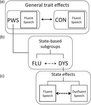
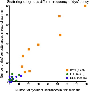
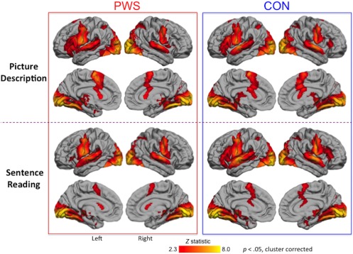
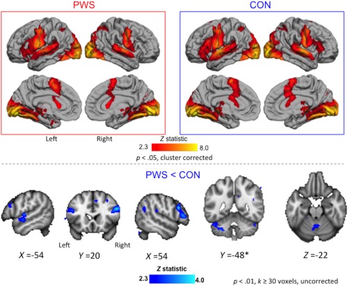
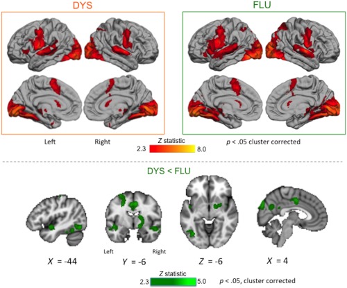
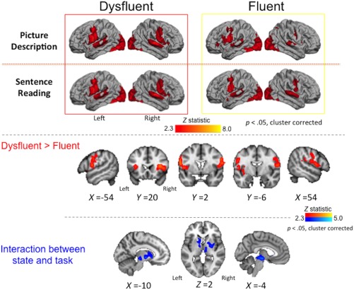
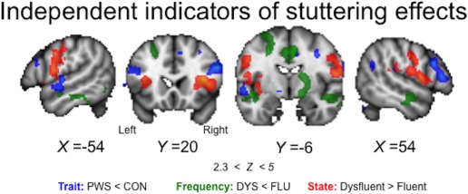
Stuttering is a disorder in which the smooth flow of speech is interrupted. People who stutter show structural and functional abnormalities in the speech and motor system. It is unclear whether functional differences reflect general traits of the disorder or are specifically related to the dysfluent speech state. We used a hierarchical approach to separate state and trait effects within stuttering. We collected sparse-sampled functional MRI during two overt speech tasks (sentence reading and picture description) in 17 people who stutter and 16 fluent controls. Separate analyses identified indicators of: (1) general traits of people who stutter; (2) frequency of dysfluent speech states in subgroups of people who stutter; and (3) the differences between fluent and dysfluent states in people who stutter. We found that reduced activation of left auditory cortex, inferior frontal cortex bilaterally, and medial cerebellum were general traits that distinguished fluent speech in people who stutter from that of controls. The stuttering subgroup with higher frequency of dysfluent states during scanning (n = 9) had reduced activation in the right subcortical grey matter, left temporo-occipital cortex, the cingulate cortex, and medial parieto-occipital cortex relative to the subgroup who were more fluent (n = 8). Finally, during dysfluent states relative to fluent ones, there was greater activation of inferior frontal and premotor cortex extending into the frontal operculum, bilaterally. The above differences were seen across both tasks. Subcortical state effects differed according to the task. Overall, our data emphasise the independence of trait and state effects in stuttering.

Keywords: basal ganglia; cerebellum; developmental stuttering; movement disorder; speech disorder.


Figures

Figure 1
Figure 1
Isolating trait and state effects in stuttering. (a) We conceptualised general traits as those things shared by people who stutter (PWS) that are different when compared to fluent controls (CON). In this way, activity during fluent speech, which occurs in both PWS and CON, can be used to isolate a general trait of stuttering. (b) In our study, about half of the PWS group overlapped with CON in the average number of dysfluent utterances per scan; the other half did not overlap with CON at all in terms of frequency of dysfluent utterances. Therefore, we used a data–driven threshold to create two subgroups of PWS: mostly fluent (FLU) and somewhat dysfluent (DYS). These subgroups were compared directly to isolate effects related to a general “proclivity” to dysfluency in the scanning environment. (c) Within the DYS subgroup, we were able to isolate specific state effects through comparing dysfluent to fluent speech epochs directly
Figure 2
Figure 2
Stuttering subgroups differed in terms of the frequency of dysfluent utterances during scan runs. This plot reflects how we determined subgroups of people who stutter using a data‐driven threshold of 10 dysfluent utterances per scan run. The mostly fluent (FLU) subgroup overlapped entirely with the fluent control group (CON) in their frequencies of dysfluent speech in the scanner. The somewhat dysfluent stuttering subgroup (DYS) did not overlap with either CON or FLU in terms of the number of dysfluent utterances across scan runs [Color figure can be viewed at http://wileyonlinelibrary.com]
Figure 3
Figure 3
Fluent speech‐related activity: People who stutter (PWS), and fluent controls (CON) activated the same network during picture description (top two rows) and sentence reading (bottom two rows). Coloured statistical maps were thresholded (Z > 2.3, p < .05) and superimposed on the lateral and medial surfaces of the left and right hemisphere using FreeSurfer [Color figure can be viewed at http://wileyonlinelibrary.com]
Figure 4
Figure 4
Trait effects during fluent speech. Averaged brain activity across both speech tasks is shown for the PWS and CON groups (boxed images, top; see legend to Figure 3 for details). Areas with reduced activity in PWS relative CON are shown in blue overlaid on sections through the MNI‐152 average brain (bottom; exploratory threshold of p < .01, k > 30 voxels, uncorrected; coordinates in MNI space). These differences were observed in the auditory cortex and lateral cerebellum of the left hemisphere, in inferior frontal gyrus bilaterally, and in the vermis of the cerebellum. *The left lateral cerebellum also showed an interaction effect with task. There were no areas where activity for PWS > CON at the exploratory threshold [Color figure can be viewed at http://wileyonlinelibrary.com]
Figure 5
Figure 5
Within‐trait effects related to stuttering subgroups. Averaged brain activity across both speech tasks is shown for the DYS and FLU subgroups (boxed images, top; see legend to Figure 3 for details). Areas with reduced activity in DYS relative to FLU subgroups across tasks are shown in green overlaid on sections through the MNI‐152 average brain (bottom; Z > 2.3, p < .05, corrected; coordinates in MNI space). These differences were observed in subcortical grey matter, cingulate cortex, left inferior temporo‐occipital cortex, superior temporal cortex bilaterally, and medial parieto‐occipital cortex. There were no areas where activity was greater in the DYS relative to the FLU subgroup [Color figure can be viewed at http://wileyonlinelibrary.com]
Figure 6
Figure 6
State effects. Averaged brain activity during each speech state (dysfluent and fluent) is shown for the picture description and sentence reading tasks separately (boxed images, top, see legend to Figure 3 for details). Areas showing greater activity during dysfluent relative to fluent states within individuals across tasks are shown in red (middle; see legend to Figure 5 for details). Individuals had greater activity during dysfluent relative to fluent speech in the lateral inferior frontal and premotor cortex extending into the opercular cortex and anterior insula bilaterally. Subcortical areas showing a significant interaction between state and task are shown in blue (bottom; see legend to Figure 5 for details) [Color figure can be viewed at http://wileyonlinelibrary.com]
Figure 7
Figure 7
Summary of general trait, specific state, and frequency effects. Areas with reduced activity in PWS relative to CON during fluent speech are shown in blue (trait effects). Areas with reduced activity in the DYS relative to the FLU subgroup of stutterers are shown in green (frequency effects). Areas with increased activity during dysfluency relative to fluency within individuals in the DYS subgroup are shown in red (state effects). The regions showing these effects are mostly spatially distinct. Coloured statistical maps are overlaid on sections through the MNI‐152 average brain (Z > 2.3, p < .05, corrected, coordinates in MNI space) [Color figure can be viewed at http://wileyonlinelibrary.com]

References

    1. Alm, P. A. (2004). Stuttering and the basal ganglia circuits: A critical review of possible relations. Journal of Communication Disorders, 37(4), 325–369. 10.1016/j.jcomdis.2004.03.001 - DOI - PubMed
    1. American Psychiatric Association , (2013). DSM‐V. American Journal of Psychiatry, 10.1176/appi.books.9780890425596.744053 - DOI
    1. Belyk, M. , Kraft, S. J. , & Brown, S. (2017). CORRIGENDUM Stuttering as a trait or state – An ALE meta‐analysis of neuroimaging studies. European Journal of Neuroscience, 45, 622–624. 10.1111/ejn.12765 - DOI - PubMed
    1. Braun, A. R. , Varga, M. , Stager, S. , Schulz, G. , Selbie, S. , Maisog, J. M. , … Ludlow, C. L. (1997). Altered patterns of cerebral activity during speech and language production in developmental stuttering. An H2(15)O positron emission tomography study. Brain, 120(5), 761–784. (Pt 5, 10.1093/brain/120.5.761 - DOI - PubMed
    1. Brown, S. , Ingham, R. J. , Ingham, J. C. , Laird, A. R. , & Fox, P. T. (2005). Stuttered and fluent speech production: An ALE meta‐analysis of functional neuroimaging studies. Human Brain Mapping, 25(1), 105–117. 10.1002/hbm.20140 - DOI - PMC - PubMed

Publication types