

Lupus nephritis: low urinary DNase I levels reflect loss of renal DNase I and may be utilized as a biomarker of disease progression
- PMID: 29624903
- PMCID: PMC6065113
- DOI: 10.1002/cjp2.99
Lupus nephritis: low urinary DNase I levels reflect loss of renal DNase I and may be utilized as a biomarker of disease progression
Abstract
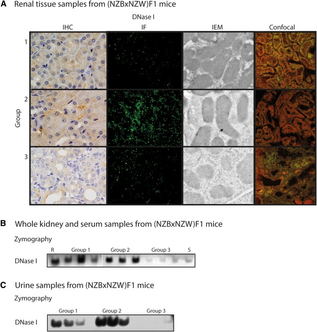
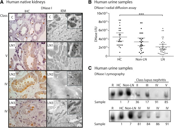
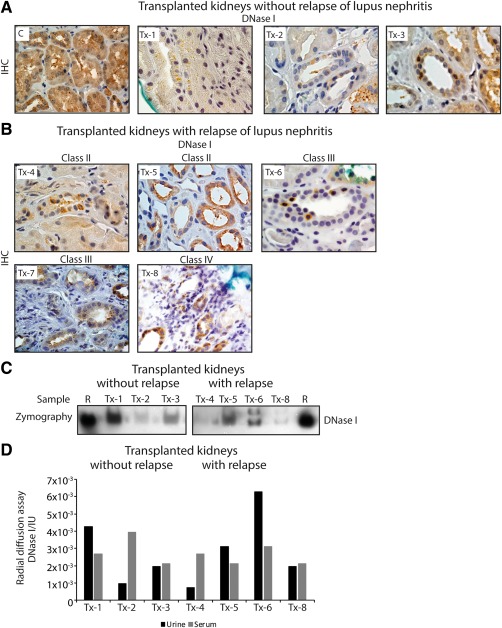
Renal DNase I is lost in advanced stages of lupus nephritis. Here, we determined if loss of renal DNase I reflects a concurrent loss of urinary DNase I, and whether absence of urinary DNase I predicts disease progression. Mouse and human DNase I protein and DNase I endonuclease activity levels were determined by western blot, gel, and radial activity assays at different stages of the murine and human forms of the disease. Cellular localization of DNase I was analyzed by immunohistochemistry, immunofluorescence, confocal microscopy, and immunoelectron microscopy. We further compared DNase I levels in human native and transplanted kidneys to determine if the disease depended on autologous renal genes, or whether the nephritic process proceeded also in transplanted kidneys. The data indicate that reduced renal DNase I expression level relates to serious progression of lupus nephritis in murine, human native, and transplanted kidneys. Notably, silencing of renal DNase I correlated with loss of DNase I endonuclease activity in the urine samples. Thus, urinary DNase I levels may therefore be used as a marker of lupus nephritis disease progression and reduce the need for renal biopsies.
Keywords: DNase I; biomarker; biopsy; lupus nephritis; urine.
© 2018 The Authors The Journal of Pathology: Clinical Research published by The Pathological Society of Great Britain and Ireland and John Wiley & Sons Ltd.
Figures
Similar articles
-
Impact of the tumor necrosis factor receptor-associated protein 1 (Trap1) on renal DNaseI shutdown and on progression of murine and human lupus nephritis.Am J Pathol. 2013 Mar;182(3):688-700. doi: 10.1016/j.ajpath.2012.11.013. Epub 2012 Dec 27. Am J Pathol. 2013. PMID: 23273922
-
Renal Dnase1 enzyme activity and protein expression is selectively shut down in murine and human membranoproliferative lupus nephritis.PLoS One. 2010 Aug 10;5(8):e12096. doi: 10.1371/journal.pone.0012096. PLoS One. 2010. PMID: 20856893 Free PMC article.
-
Acquired loss of renal nuclease activity is restricted to DNaseI and is an organ-selective feature in murine lupus nephritis.Am J Pathol. 2011 Sep;179(3):1120-8. doi: 10.1016/j.ajpath.2011.05.011. Epub 2011 Jun 30. Am J Pathol. 2011. PMID: 21723244 Free PMC article.
-
Murine and Human Lupus Nephritis: Pathogenic Mechanisms and Theoretical Strategies for Therapy.Semin Nephrol. 2015 Sep;35(5):427-38. doi: 10.1016/j.semnephrol.2015.08.004. Semin Nephrol. 2015. PMID: 26573545 Review.
-
Nuclease deficiencies promote end-stage lupus nephritis but not nephritogenic autoimmunity in (NZB × NZW) F1 mice.Immunol Cell Biol. 2011 Jan;89(1):90-9. doi: 10.1038/icb.2010.75. Epub 2010 Jun 15. Immunol Cell Biol. 2011. PMID: 20548325 Review.
Cited by
-
The greatest contribution to medical science is the transformation from studying symptoms to studying their causes-the unrelenting legacy of Robert Koch and Louis Pasteur-and a causality perspective to approach a definition of SLE.Front Immunol. 2024 Feb 1;15:1346619. doi: 10.3389/fimmu.2024.1346619. eCollection 2024. Front Immunol. 2024. PMID: 38361929 Free PMC article.
-
Why is it so difficult to understand why we don't understand human systemic lupus erythematosus? Contemplating facts, conflicts, and impact of "the causality cascade paradigm".Front Immunol. 2025 Jan 28;15:1507792. doi: 10.3389/fimmu.2024.1507792. eCollection 2024. Front Immunol. 2025. PMID: 39936150 Free PMC article. Review.
-
Expanding the Toolbox for Label-Free Enzyme Assays: A Dinuclear Platinum(II) Complex/DNA Ensemble with Switchable Near-IR Emission.Molecules. 2019 Dec 1;24(23):4390. doi: 10.3390/molecules24234390. Molecules. 2019. PMID: 31805648 Free PMC article.
-
Multimodal insights into adult neurogenesis: An integrative review of multi-omics approaches.Heliyon. 2025 Feb 13;11(4):e42668. doi: 10.1016/j.heliyon.2025.e42668. eCollection 2025 Feb 28. Heliyon. 2025. PMID: 40051854 Free PMC article. Review.
-
Leveraging Heterogeneity in Systemic Lupus Erythematosus for New Therapies.Trends Mol Med. 2021 Feb;27(2):152-171. doi: 10.1016/j.molmed.2020.09.009. Epub 2020 Oct 9. Trends Mol Med. 2021. PMID: 33046407 Free PMC article. Review.
References
-
- Davidson A, Bethunaickan R, Berthier C, et al Molecular studies of lupus nephritis kidneys. Immunol Res 2015; 63: 187–196. - PubMed
-
- Zubair A, Frieri M. Lupus nephritis: review of the literature. Curr Allergy Asthma Rep 2013; 13: 580–586. - PubMed
-
- Hogan J, Appel GB. Update on the treatment of lupus nephritis. Curr Opin Nephrol Hypertens 2013; 22: 224–230. - PubMed
Publication types
MeSH terms
Substances
LinkOut - more resources
Full Text Sources
Other Literature Sources
