
Perspective on Oncogenic Processes at the End of the Beginning of Cancer Genomics

Li Ding et al. Cell. .

Abstract

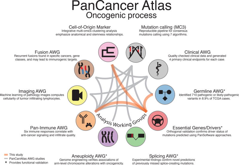
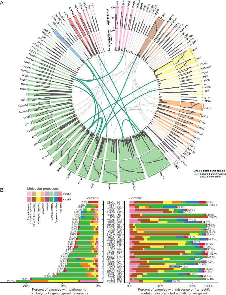
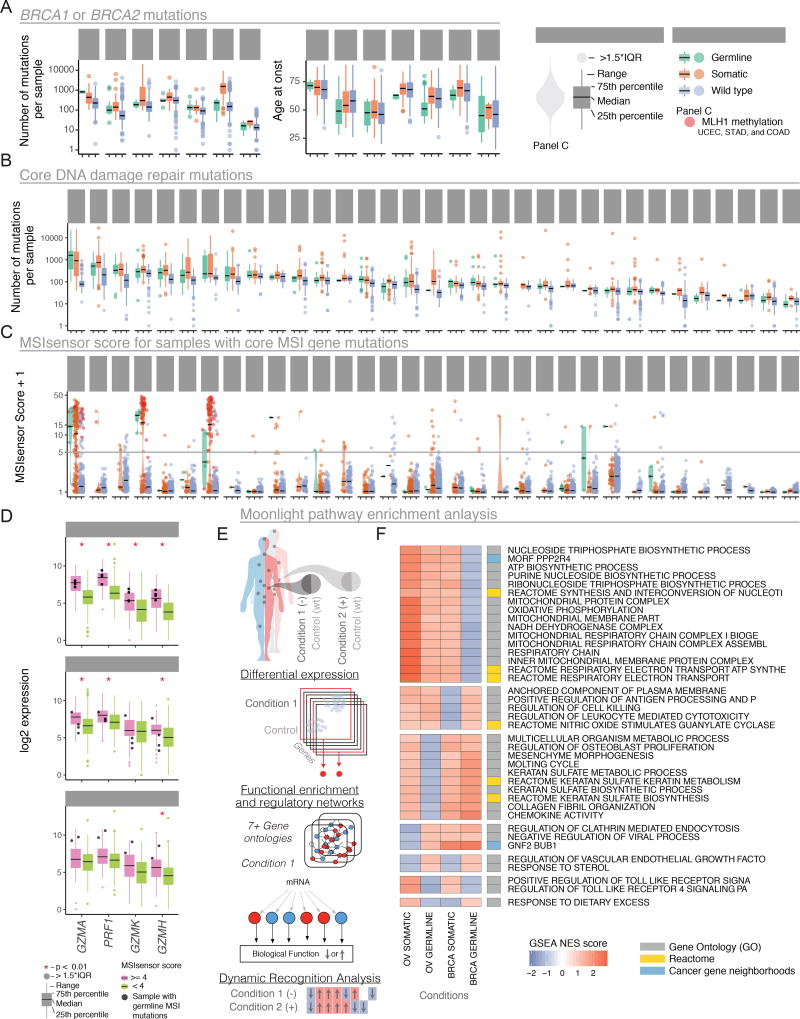
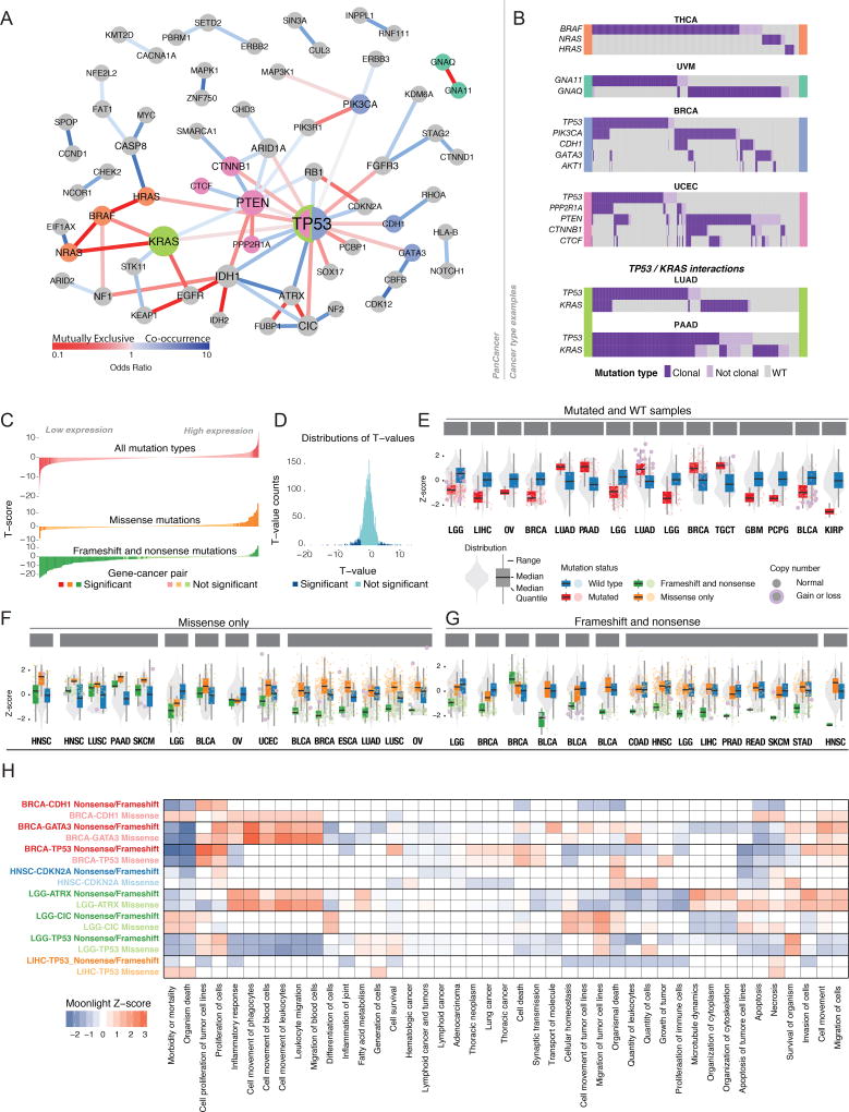
The Cancer Genome Atlas (TCGA) has catalyzed systematic characterization of diverse genomic alterations underlying human cancers. At this historic junction marking the completion of genomic characterization of over 11,000 tumors from 33 cancer types, we present our current understanding of the molecular processes governing oncogenesis. We illustrate our insights into cancer through synthesis of the findings of the TCGA PanCancer Atlas project on three facets of oncogenesis: (1) somatic driver mutations, germline pathogenic variants, and their interactions in the tumor; (2) the influence of the tumor genome and epigenome on transcriptome and proteome; and (3) the relationship between tumor and the microenvironment, including implications for drugs targeting driver events and immunotherapies. These results will anchor future characterization of rare and common tumor types, primary and relapsed tumors, and cancers across ancestry groups and will guide the deployment of clinical genomic sequencing.

Keywords: TCGA; cancer; cancer genomics; omics; oncogenic process.


Figures

Figure 1
Figure 1. Overview of the PanCancer Atlas oncogenic process group
PanCan Atlas studies use data from multiple working groups, with relationships shown by gray edges between associated studies. New connections described in this study are shown as orange edges.
Figure 2
Figure 2. Sequence level evaluation of samples with pathogenic germline mutations
A Circos plot for each predisposition cancer gene. Width of each slice is proportional to germline variant frequency. The outermost tier shows age at onset, while middle indicates total number of somatic mutations for each sample. Links designate one sample that has multiple pathogenic or likely pathogenic germline mutations and are green if one of the genes is from the Fanconi anemia pathway. B shows somatic and germline driver genes grouped into 8 molecular process categories. On the x-axis, germline and somatic proportions are plotted using number of samples as the denominator. Cancers are sorted by increasing germline contribution.
Figure 3
Figure 3. Evaluation of BRCA1/BRCA2 DDR, and MSI genes using somatic and germline variation
A Samples with BRCA1 or BRCA2 mutations are grouped by cancer type and stratified by somatic, germline, or wild-type status. Box-plots highlight mutations per-sample (left) and age at onset (right). Outlier samples are plotted as points. B Box-plots for samples having mutations in DNA damage response genes grouped by cancer. C Violin plots of MSIsensor scores with samples grouped based on mutation status of MSI genes. Samples with MLH1 promoter methylations status are shown in red. D Gene expression differences for cytokine activators for three cancer types. Black dots are samples with predisposition germline mutation in MSI genes. Red stars highlight significant differences between groups. E Moonlight workflow shows how samples were stratified based on germline vs. wild type (condition 1) and somatic vs. wild type (condition 2) and integrated across pathways with genes that are labeled as differentially expressed. These were then compared using dynamic recognition analysis to identify patterns. F Normalized scores from gene set enrichment analysis for germline and somatic mutations in BRCA1 and/or BRCA2 only, as conditions of OV and BRCA cancer types. Only the first 50 characters of each pathway are shown (additional information in Supplemental Figure 1).
Figure 4
Figure 4. Interactions between somatic driver events
A Mutual exclusivity and cooccurrence of driver events. Nodes sized according to degree and edges colored according to odds ratio of pairs of drivers: red for mutually exclusive (OR < 1) and blue for co-occurrence (OR > 1). B Tissue-specific interactions of driver events. Waterfall plots show whether each patient has clonal (dark purple), sub-clonal (light purple), or no driver mutation (gray). Each plot is flanked with a color corresponding to genes in panel A. C Landscape of cis-expression changes shown for three mutation types, with FDR < 0.1 considered significant. D Distribution of T-values for gene expression analyses. ECis-effects of mutations in expression of driver genes. Gray violin plot depicts expression in all samples of driver gene in the tissue marked below each plot. Red boxes show expression of samples with any mutations in that gene blue boxes show expression for samples with no mutation in that gene. Each dot represents a sample and is red if there is a copy number alteration of the gene. F Same information as in E, but separating samples according to frameshift and nonsense (green) versus missense mutations (orange). Selected genes show the top-15 t-values when comparing between the missense and no-mutation groups (FDR < 0.1). G Same as in F, but genes selected by top-15 t-values between nonsense/frameshift and no-mutations groups. H Moonlight scores for groups of mutations in driver genes in specific cancer types (y-axis) and genes annotated with several GO terms (x-axis). Boxes colored red or blue if Moonlight Z-score is positive (overexpression of the biological function) or negative (downregulation), respectively. See also supplemental figure 2.
Figure 5
Figure 5. Relationships between oncogenic processes and driver genes
A Identifying processes deregulated by driver gene modules using OncoIMPACT. Pathways associated with each module were identified using enrichment analysis (Methods). B Relationships among oncogenic processes, cancer types, and driver genes. (Left) Heatmap shows fraction of samples with deregulated processes associated with sample-specific driver mutations. The three most frequently mutated driver genes are shown with each cancer type. (Right) Graph of associations between processes and top three genes predicted to be responsible for their deregulation. Grey cells represent non-significant fraction of patients (binomial test, p-value Bonferroni corrected > 0.05). Edge widths represent relative fraction of samples with deregulated processes associated to each driver gene. C Oncoprint of mutational profile of the 5 most mutated genes associated with deregulation of 3 biological processes. (Left) Different samples harbor driver genes in a mutually exclusive manner, suggesting many samples have only one process driver gene. (Right) Number of samples having driver gene mutated. P-values are computed using R-exclusivity test (Methods).
Figure 6
Figure 6. Complexities of multi dimensional molecular evaluation
A Clustering analysis was performed using 3 substrates: methylation, mRNA, and RPPA. Samples divided into 24 methylation clusters, 41 mRNA, and 10 RPPA clusters. Links show each tumor was given a unique cluster combination identifier. B Gene enrichment analysis for each cluster assignment is displayed as a volcano plot. Dashed square is enlarged in an inset. Overlapping dots show number of samples in the cluster assignment (dark blue) and the number of samples with a given mutation superimposed (light blue), jointly indicating the mutated proportion in that cluster. C The 21 most gene enriched cluster identities, with breakdown by tissue type proportion and most frequently mutated gene from that cluster identity. Sample size for each identity appears in bar plot. D The 58 cluster identities having ≥20 samples. Pie chart illustrates fraction of uniform clusters, where 90% of samples within a cluster are from a single cancer type.
Figure 7
Figure 7. Statistical associations and predicted interactions within the tumor microenvironment
A Networks of driver gene events in distinct cancer immune subtypes C1-C6 shown in each subpanel. Lines between events and immune cells are green if correlation between immune cell in samples with the driver event is positive and red if negative. Lines between cell types, ligands, and receptors denote interaction pairs known to occur in other contexts and for which there are concordant values across multiple tumor samples in the subtype. B Heatmap shows Spearman correlation between number of predicted neoantigens in each sample of each immune subtype and proportion of different types of immune cells. Colored outline boxes are detailed in the next panel. C In subtypes C1 and C2, proportion of CD8 T cells increases with burden of predicted neoantigens (left two plots). Correlation between number of neoantigens and Neutrophils in samples of C3 subtype (top right) and between number of neoantigens and fraction of macrophages in the TME in samples with C5 immune response (bottom right).

References

    1. Akbani R, Akdemir KC, Aksoy BA, Albert M, Ally A, Amin SB, Arachchi H, Arora A, Auman JT, Ayala B. Genomic classification of cutaneous melanoma. Cell. 2015;161:1681–1696. - PMC - PubMed
    1. Alvarez MJ, Shen Y, Giorgi FM, Lachmann A, Ding BB, Ye BH, Califano A. Functional characterization of somatic mutations in cancer using network-based inference of protein activity. Nat Genet. 2016;48:838–847. - PMC - PubMed
    1. Bashashati A, Haffari G, Ding J, Ha G, Lui K, Rosner J, Huntsman DG, Caldas C, Aparicio SA, Shah SP. DriverNet: uncovering the impact of somatic driver mutations on transcriptional networks in cancer. Genome biology. 2012;13:R124. - PMC - PubMed
    1. Bassi R, Giussani P, Anelli V, Colleoni T, Pedrazzi M, Patrone M, Viani P, Sparatore B, Melloni E, Riboni L. HMGB1 as an autocrine stimulus in human T98G glioblastoma cells: role in cell growth and migration. J Neurooncol. 2008;87:23–33. - PubMed
    1. Bertrand D, Chng KR, Sherbaf FG, Kiesel A, Chia BK, Sia YY, Huang SK, Hoon DS, Liu ET, Hillmer A. Patient-specific driver gene prediction and risk assessment through integrated network analysis of cancer omics profiles. Nucleic acids research. 2015;43:e44–e44. - PMC - PubMed

Publication types