. 2018 Apr 17;48(4):716-729.e8.
doi: 10.1016/j.immuni.2018.03.015. Epub 2018 Apr 3.

KLRG1+ Effector CD8+ T Cells Lose KLRG1, Differentiate into All Memory T Cell Lineages, and Convey Enhanced Protective Immunity

Affiliations

KLRG1+ Effector CD8+ T Cells Lose KLRG1, Differentiate into All Memory T Cell Lineages, and Convey Enhanced Protective Immunity

Dietmar Herndler-Brandstetter et al. Immunity. .

Abstract

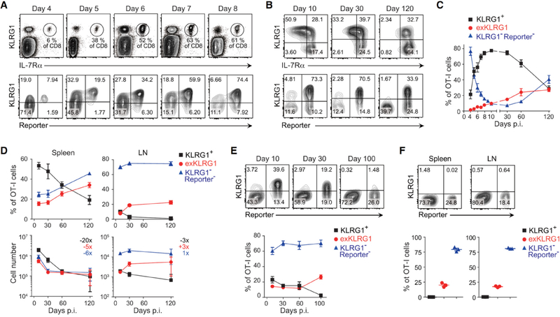
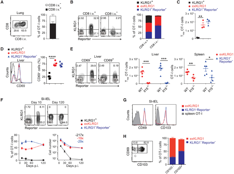
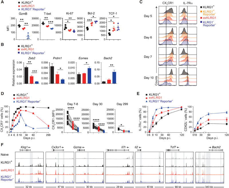
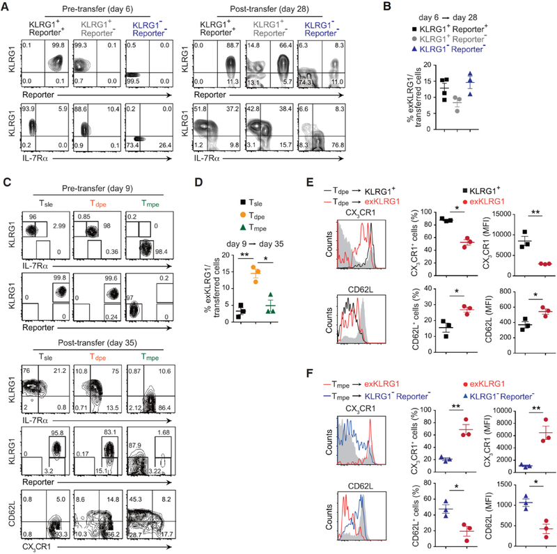
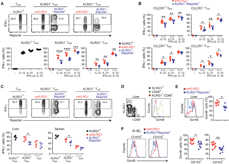
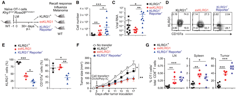
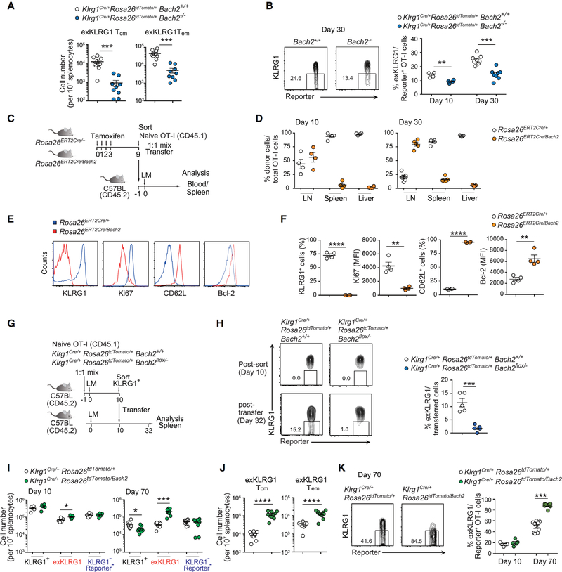
Protective immunity against pathogens depends on the efficient generation of functionally diverse effector and memory T lymphocytes. However, whether plasticity during effector-to-memory CD8+ T cell differentiation affects memory lineage specification and functional versatility remains unclear. Using genetic fate mapping analysis of highly cytotoxic KLRG1+ effector CD8+ T cells, we demonstrated that KLRG1+ cells receiving intermediate amounts of activating and inflammatory signals downregulated KLRG1 during the contraction phase in a Bach2-dependent manner and differentiated into all memory T cell linages, including CX3CR1int peripheral memory cells and tissue-resident memory cells. "ExKLRG1" memory cells retained high cytotoxic and proliferative capacity distinct from other populations, which contributed to effective anti-influenza and anti-tumor immunity. Our work demonstrates that developmental plasticity of KLRG1+ effector CD8+ T cells is important in promoting functionally versatile memory cells and long-term protective immunity.

Keywords: Bach2; CD8 T cell; CX(3)CR1; cancer; fate mapping; inflammation; influenza; memory; plasticity; tissue-resident.


Conflict of interest statement

DECLARATION OF INTERESTS

The authors declare no competing interests.

Figures

Figure 1.
Figure 1.. Effector CD8+ T Cells Lose KLRG1 Expression and Differentiate into Long-Lived Memory Cells.
(A and B) Expression of KLRG1 and fate mapping in effector OT-I cells in the blood following LM infection. (C) Frequency of KLRG1+, exKLRG1 and KLRG1 Reporter cells among OT-I cells in the blood up to 120 days p.i. with LM. (D) Percentage (top) and number (bottom) of OT-I cell subsets in the spleen and LN following LM infection. The numbers indicate fold difference in cell number between days 10 and 120. (E) Frequency of KLRG1+, exKLRG1 and KLRG1 Reporter cells among OT-I cells in the blood up to 100 days p.i. with VSV. (F) Percentage of KLRG1+, exKLRG1 and KLRG1 Reporter cells among OT-I cells in the spleen and LN 100 days p.i. with VSV. Mean ± SEM are shown. Data are pooled from 2–4 independent experiments with 4–12 (C) or 3–12 mice per time point (D), or are representative of 2–3 independent experiments with 3–5 mice per time point (A, E, F). See also Figure S1 and S2.
Figure 2.
Figure 2.. KLRG1+ Effector CD8+ T Cells Differentiate into Long-Lived Trm Cells.
(A-C) Frequency and number of resident (i.v.) and circulating (i.v.+) memory OT-I cell subsets in the lung 60 days p.i. with LM. Circulating cells were identified by i.v. injection of anti-CD8α antibody. (D) Frequency of CD69+ memory OT-I cell subsets in the liver 62 days p.i. with LM. (E) Frequency and number of CD69+ Trm cells in the liver 30 days p.i. with LM. (F) Frequency and number of OT-I cell subsets in the intraepithelial lymphocyte fraction of the small intestine (SI-IEL) 10 and 120 days p.i. with LM. Numbers indicate fold difference in cell number between days 10 and 120. (G) Expression of CD69 and CD103 in OT-I cell subsets in the SI-IEL of mice orally infected with LM (day 42 p.i.). (H) Frequency of CD103 CD69+ or CD103+ CD69+ memory OT-I cell subsets in the SI-IEL. Mean ± SEM are shown. ** P < 0.01, *** P < 0.001 and **** P < 0.0001 (unpaired two-tailed Student’s t-test). Data are representative of two independent experiments with 3 (A-C), 4–5 (D, E), or 8 mice (G, H), or are pooled from 2–3 independent experiments with 4–9 mice per time point (F). See also Movies S1 and S2.
Figure 3.
Figure 3.. ExKLRG1 Effector CD8+ T cells Express Cytotoxicity, Survival, and Proliferation Molecules at an Intermediate Level.
(A) Expression of GzmB, T-bet, Ki-67, Bcl-2, and TCF-1 in splenic effector OT-I cell subsets 9–10 days p.i. with LM. (B) Expression of effector and memory signature genes in splenic OT-I cell subsets 8–11 days p.i. with LM. (C-E) Time-dependent expression of CX3CR1 and IL-7Rα in OT-I cell subsets in the blood following LM infection. (F) Normalized ATAC-seq signal profiles across 7 gene loci in splenic naïve and effector OT-I cell subsets (8 days p.i. with LM). Peaks differentially expressed between OT-I cell subsets are highlighted in grey. Mean ± SEM are shown. * P < 0.05, ** P < 0.01 and *** P < 0.001 (unpaired two-tailed Student’s t-test). Data are representative of 2–3 independent experiments with 4–8 mice (A, C), pooled from 2–3 independent experiments with 3–11 mice per time point (B, D, E), or 2 independent experiments with pooled cells from 2–3 mice (F). See also Figure S3.
Figure 4.
Figure 4.. Developmental Plasticity of Effector CD8+ T cell Subsets Following LM Infection.
(A) Expression of KLRG1, IL-7Rα and tdTomato in splenic effector OT-I cell subsets 6 days p.i. with LM (pre-transfer) and 22 days post-transfer (day 28 p.i.). (B) Development of exKLRG1 memory cells (day 28 p.i.) from three different effector OT-I cell subsets 6 days p.i. with LM. (C) Expression of KLRG1, IL-7Rα, tdTomato, CD62L and CX3CR1 in splenic effector OT-I cell subsets 9 days p.i. with LM (pre-transfer) and 26 days post-transfer (day 35 p.i.). (D) Development of exKLRG1 memory cells (day 35 p.i.) from three different effector OT-I cell subsets 9 days p.i. with LM. (E) Expression of CX3CR1 and CD62L in KLRG1+ and exKLRG1 memory cells 26 days post transfer of day 9 Tdpe cells. Host CD8+ T cells served as a control (gray line). (F) Expression of CX3CR1 and CD62L in exKLRG1 and KLRG1 Reporter memory cells 26 days post transfer of day 9 Tmpe cells. Host CD8+ T cells served as a control (gray line). Mean ± SEM are shown. * P < 0.05 and ** P < 0.01 (unpaired two-tailed Student’s t-test). Data are representative of two independent experiments with 3–4 mice. See also Figure S4.
Figure 5.
Figure 5.. ExKLRG1 Memory Cell Subsets Retain High Cytotoxic Capacity and Responsiveness to IL-12.
(A and B) Production of IFN-γ in splenic Tem (KLRG1+ CD62L or KLRG1 CD62L) and Tcm (KLRG1 CD62L+) OT-I cells (A) or Tem (KLRG1 CX3CR1+ CD62L) and Tcm (KLRG1 CX3CR1+ CD62L+) OT-I cells (B) following stimulation with IL-12 (density plots), IL-12 + IFNα/β or IL-12 + IL-15 for 7 hours in vitro. (C) Memory OT-I cells were generated as described in Figure S1E and mice were challenged 90 days later with Listeria monocytogenes, which did not express OVA. Production of IFN-γ in Tem (KLRG1+ CD62L or KLRG1 CD62L) and Tem (KLRG1 CD62L+) OT-I cells in spleen and liver (density plots) 12 hours after rechallenge. (D) Expression of GzmB in circulating memory (KLRG1+CD69 or KLRG1CD69) and Trm (KLRG1CD69+) cells within the endogenous OVA-tetramer+ CD44hi CD8+ T cell population in the liver 108 days p.i. with LM. (E) Expression of GzmB in KLRG1 Reporter Trm and exKLRG1 tetramer+ Trm cells in the liver 108 days p.i. with LM. (F) Expression of GzmB in KLRG1 Reporter Trm and exKLRG1 Trm cells within intraepithelial CD103+ CD69+ and CD103 CD69+ OT-I cell subsets 42 days after oral infection with LM (n=8). Mean ± SEM are shown. * P < 0.05, ** P < 0.01, *** P < 0.001 and **** P < 0.0001 (unpaired two-tailed Student’s t-test). The data are representative of two (C-F) or three (A, B) independent experiment with 3–7 mice. See also Figure S5.
Figure 6.
Figure 6.. ExKLRG1 Memory CD8+ T cells Mount Potent Anti-Influenza and Anti-Tumor Responses.
(A) Schematic of the adoptive transfer and infection experiments. (B) Mice receiving memory OT-I cell subsets were challenged i.n. with OVA-expressing influenza virus (FLU). Seven days later, the number of effector OT-I cells in the lung was determined. (C) Viral RNA in the lung 7 days p.i. with FLU was determined by quantitative RT-PCR. (D and E) Expression of KLRG1 and CD107a in effector OT-I cell subsets in the lung 7 days p.i. with FLU. (F) Tumor size of mice s.c. injected with melanoma cells (B16-OVA) followed by adoptive transfer of memory OT-I cell subsets. (G) Percentage of transferred OT-I cells within total CD8+ T cells in the LN, spleen and tumor at day 17 after tumor inoculation. Data were analyzed by unpaired two-tailed Student’s t-test (B, C, E) or two-way ANOVA
(F). Mean ± SEM are shown. * P < 0.05, ** P < 0.01 and *** P < 0.001. Data are pooled from 2–3 experiments with 10–11 mice (B and C), or representative of two (D, E) or three (F, G) independent experiments with 3–4 (D, E) and 5–6 (F, G) mice.
Figure 7.
Figure 7.. Bach2 Supports the Differentiation of KLRG1+ Effector Cells into ExKLRG1 Memory Cells.
(A) Naive Klrg1Cre/+Rosa26tdTomato/+Bach2+/+ and Klrg1Cre/+Rosa26tdTomato/+Bach2−/− OT-I cells from different CD45 congenic backgrounds were co-transferred into WT mice, followed by infection with LM one day later. The number of exKLRG1 Tcm and exKLRG1 Tem OT-I cells in the spleen 30 days p.i. with LM is shown. (B) Percentage of exKLRG1 cells among Reporter+ donor OT-I cells in the spleen 10 and 30 days p.i. with LM. (C) Schematic of the experimental protocol for (D-F). Tamoxifen was injected for four consecutive days to induce Cre-dependent Bach2 expression in OT-I Rosa26ERT2Cre/Bach2 mice. After 9 days, Bach2-expressed GFP+ naïve OT-I cells were sorted, mixed with Rosa26ERT2Cre/+ naïve OT-I cells at a 1:1 ratio, and transferred into C57BL/6 mice followed by infection with LM one day later. (D) Relative frequency of Rosa26ERT2Cre/+ and Rosa26ERT2Cre/Bach OT-I cells 10 and 30 days p.i. with LM in the LN, spleen and liver. (E and F) Expression of CD62L, KLRG1, Ki-67 and Bcl-2 in Rosa26ERT2Cre/+ and Rosa26ERT2Cre/Bach OT-I cells 10 days p.i. with LM. (G) Schematic of the experimental protocol for (H). (H) Expression of KLRG1 and Reporter in OT-I cells post-transfer of day 10 KLRG1+ effector OT-I cells into infection-matched WT mice (32 days p.i. with LM). (I) Naive Klrg1Cre/+Rosa26tdTomato/+ and Klrg1Cre/+Rosa26tdTomato/Bach2 OT-I cells from different CD45 congenic backgrounds were co-transferred into WT mice, followed by infection with LM one day later. The number of KLRG1+, exKLRG1 and KLRG1Reporter OT-I cells in the spleen at days 10 and 70 p.i. with LM is shown. (J) Percentage of exKLRG1 cells among Reporter+ OT-I cells in the spleen 70 days p.i. with LM. (K) Number of exKLRG1 Tcm and exKLRG1 Tem OT-I cells in the spleen 70 days p.i. with LM. Mean ± SEM are shown. * P < 0.05, ** P < 0.01, *** P < 0.001, **** P < 0.0001 (unpaired two-tailed Student’s t-test). Data are representative of two independent experiments with 5 (D-H) or 4–10 (A, B, I-K) mice. See also Figure S5, S6, and S7.

Comment in

References

    1. Anderson KG, Mayer-Barber K, Sung H, Beura L, James BR, Taylor JJ, Qunaj L, Griffith TS, Vezys V, Barber DL, and Masopust D (2014). Intravascular staining for discrimination of vascular and tissue leukocytes. Nat Protoc 9, 209–222. - PMC - PubMed
    1. Araki K, Turner AP, Shaffer VO, Gangappa S, Keller SA, Bachmann MF, Larsen CP, and Ahmed R (2009). mTOR regulates memory CD8 T-cell differentiation. Nature 460, 108–112. - PMC - PubMed
    1. Bergsbaken T, and Bevan MJ (2015). Proinflammatory microenvironments within the intestine regulate the differentiation of tissue-resident CD8+ T cells responding to infection. Nat Immunol 16, 406–414. - PMC - PubMed
    1. Best JA, Blair DA, Knell J, Yang E, Mayya V, Doedens A, Dustin ML, Goldrath AW, and Immunological Genome Project C (2013). Transcriptional insights into the CD8+ T cell response to infection and memory T cell formation. Nat Immunol 14, 404–412. - PMC - PubMed
    1. Bottcher JP, Beyer M, Meissner F, Abdullah Z, Sander J, Hochst B, Eickhoff S, Rieckmann JC, Russo C, Bauer T, et al. (2015). Functional classification of memory CD8+ T cells by CX3CR1 expression. Nat Commun 6, 8306. - PMC - PubMed

Publication types

MeSH terms