. 2018 Jun;46(6):860-864.
doi: 10.1124/dmd.118.080838. Epub 2018 Apr 6.

Sulfotransferase 4A1 Increases Its Expression in Mouse Neurons as They Mature

Affiliations

Sulfotransferase 4A1 Increases Its Expression in Mouse Neurons as They Mature

Takuyu Hashiguchi et al. Drug Metab Dispos. 2018 Jun.

Abstract

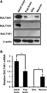
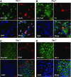
Cytosolic sulfotransferases (SULTs) catalyze sulfation and play essential roles in detoxification of xenobiotics as well as inactivation of endobiotics. SULT4A1, which was originally isolated as a brain-specific sulfotransferase, is the most highly conserved isoform among SULTs in vertebrates. Here, expression of SULT4A1 was examined neuron enriched and neuron-glia mixed cells derived from mouse embryo brains at day 14 gestation and mixed glia from 2-day-old neonate brains. Western blots showed an increase of SULT4A1 expression as neurons maturated. Reverse-transcription polymerase chain reaction and agarose gel analysis found two different forms (variant and wild type) of SULT4A1 mRNA in neurons; the level of wild type correlated with the protein level of SULT4A1. SULT1E1 was not expressed in mouse brains, neuron-enriched cells, or mixed glia cells. SULT1A1 protein was only detected in adult brains. Immunofluorescence staining of neuron-glia mixed cells confirmed selective expression of SULT4A1 in neurons, including dopaminergic neurons, but not in either astrocytes or microglia. Thus, SULT4A1 is a neuron-specific sulfotransferase and may play a role in neuronal development.


Figures

Fig. 1.
Fig. 1.
Neuron-specific expression of SULT4A1. (A) Western blots. SULT expressions were examined in 8-week-old adult mouse brains, 2-day-old neonate brains, mixed glia cells, and neuron-enriched cells using an anti-SULT4A1, SULT1A1, or SULT1E1 antibody. Sample preparations were independently conducted three times, and the representative Western blot image was indicated. (B) Real-time PCR. SULT4A1 mRNA levels using the tissues or cells as described in (A). Each value is shown as the mean ± S.D. (n = 3). Asterisks indicate significant differences between experimental groups (*P < 0.05).
Fig. 2.
Fig. 2.
SULT4A1 expression during neuron maturation. (A) Mesencephalic neuron-glia cells were cultured as described in the Materials and Methods section. Western blots were performed as described in Fig. 1. A representative image of three independent experiments was indicated. (B) Real-time PCR. Total RNAs were extracted from each stage of neuron glia cultures. (C) SULT4A1 mRNAs were amplified by reverse-transcription PCR. (D) Real-time PCR. SULT2B1 mRNAs were amplified as indicated in (B). Each value is shown as the mean ± S.D. (n = 3). Asterisks indicate significant differences between experimental groups (*P < 0.05; **P < 0.01; ***P < 0.001; ****P < 0.0001). WT, wild type.
Fig. 3.
Fig. 3.
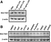
Effects of various compounds on SULT4A1 expression in mouse primary neurons. (A) Mouse neuron-enriched cells were treated with lipopolysaccharide (LPS; 1–15 μg/ml) for 24 hours and harvested for Western blot analysis. (B) The cells were treated with epidermal growth factor (EGF; 10 ng/ml), hepatocyte growth factor (HGF; 50 ng/ml), phenobarbital (PB; 1 mM), CITCO (1 μM), rifampicin (RIF; 10 μM), GW4064 (5 μM), forskolin (10 μM), 3,3′,5-triiodo-l-thyronine (T3; 10 nM), pregnenolone (PREG; 500 nM), DHEA (100 nM), or DHEA-sulfate (DHEAS; 100 nM) for 24 hours, followed by Western blot analysis. A representative Western blot image of three independent experiments was indicated. DMSO, dimethylsulfoxide.
Fig. 4.
Fig. 4.
Double fluorescence staining of mouse primary neuron/glia cells. Mesencephalic primary neuron-glia cells were prepared from E14 mouse embryos and cultured for 7 days. Cells were double stained by an anti-SULT4A1 antibody with NeuN (A), TH (B), glial fibrillary acidic protein (GFAP) (C), or IBA-1 (D). Nuclei was stained by 4′,6-diamidino-2-phenylindole (DAPI).

References

    1. Allali-Hassani A, Pan PW, Dombrovski L, Najmanovich R, Tempel W, Dong A, Loppnau P, Martin F, Thornton J, Edwards AM, et al. (2007) Structural and chemical profiling of the human cytosolic sulfotransferases [published correction appears in PLoS Biol (2007) 5:e165]. PLoS Biol 5:e97. - PMC - PubMed
    1. Block ML, Hong JS. (2005) Microglia and inflammation-mediated neurodegeneration: multiple triggers with a common mechanism. Prog Neurobiol 76:77–98. - PubMed
    1. Cepeda C, Murphy KP, Parent M, Levine MS. (2014) The role of dopamine in Huntington’s disease. Prog Brain Res 211:235–254. - PMC - PubMed
    1. Chen SH, Oyarzabal EA, Hong JS. (2013) Preparation of rodent primary cultures for neuron-glia, mixed glia, enriched microglia, and reconstituted cultures with microglia. Methods Mol Biol 1041:231–240. - PMC - PubMed
    1. Falany CN, Krasnykh V, Falany JL. (1995) Bacterial expression and characterization of a cDNA for human liver estrogen sulfotransferase. J Steroid Biochem Mol Biol 52:529–539. - PubMed

Publication types