

A Tumor-Imaging Method Targeting Cancer-Associated Fibroblasts
- PMID: 29626120
- PMCID: PMC6126438
- DOI: 10.2967/jnumed.118.210435
A Tumor-Imaging Method Targeting Cancer-Associated Fibroblasts
Abstract
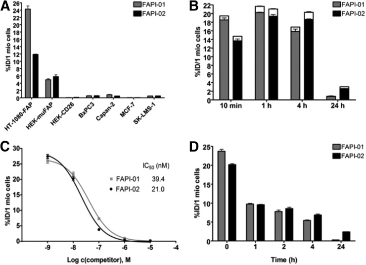
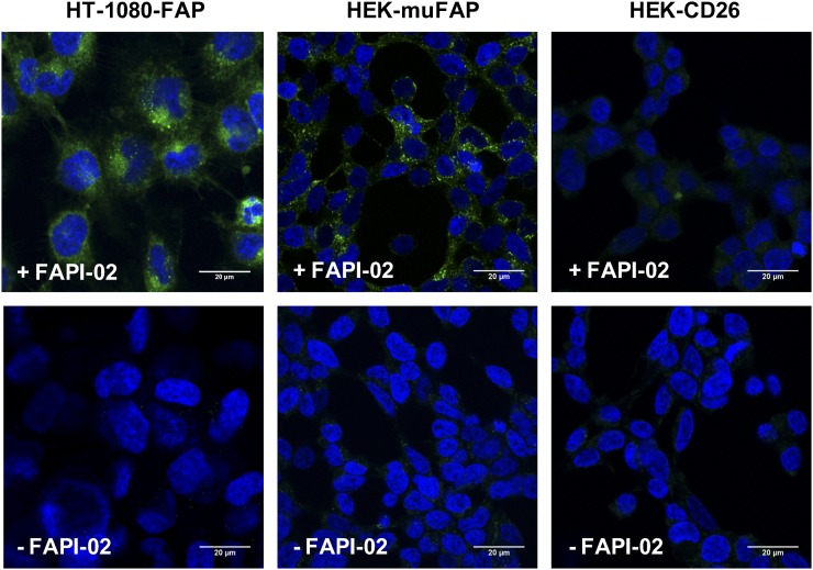
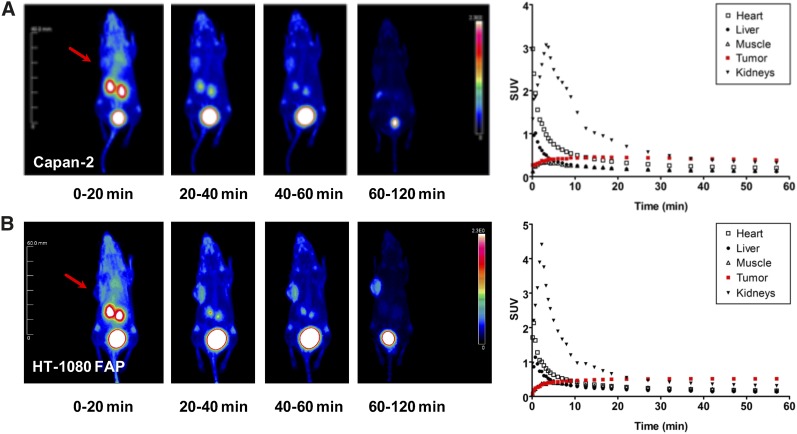
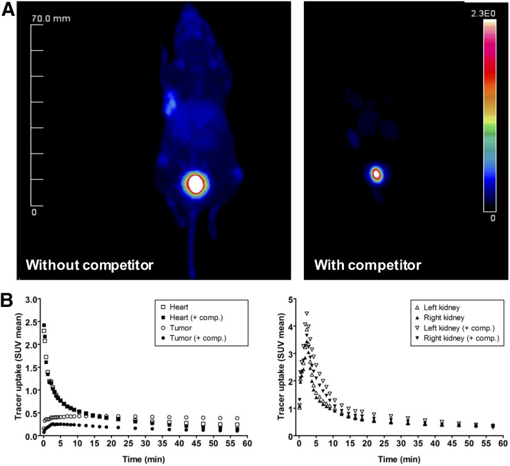
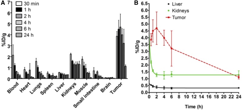
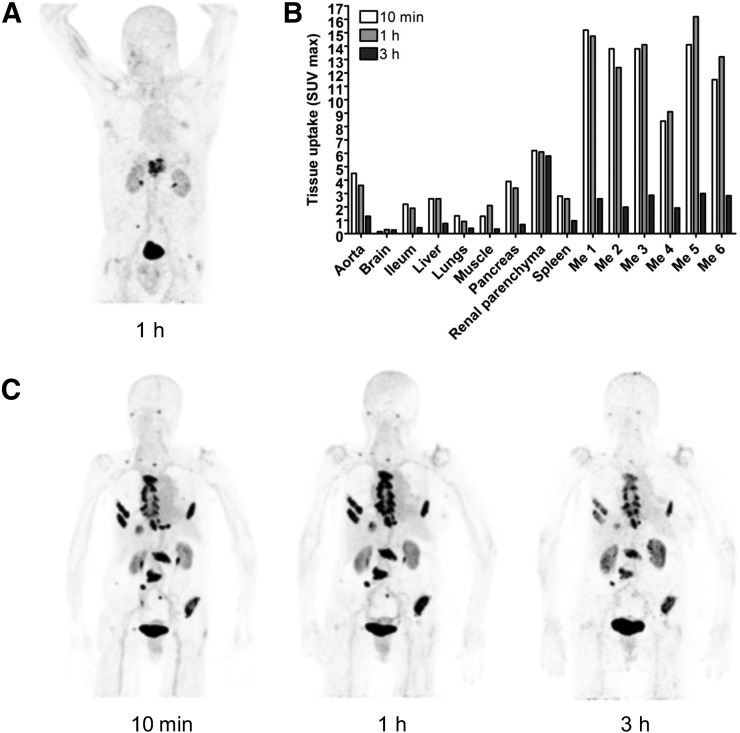
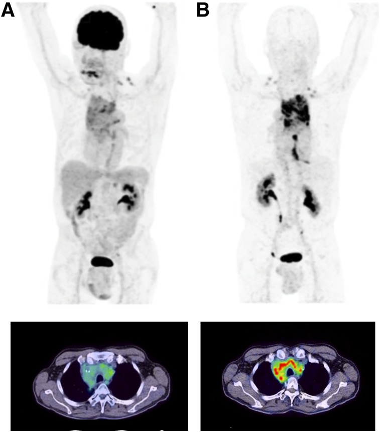
The tumor stroma, which accounts for a large part of the tumor mass, represents an attractive target for the delivery of diagnostic and therapeutic compounds. Here, the focus is notably on a subpopulation of stromal cells, known as cancer-associated fibroblasts, which are present in more than 90% of epithelial carcinomas, including pancreatic, colon, and breast cancer. Cancer-associated fibroblasts feature high expression of fibroblast activation protein (FAP), which is not detectable in adult normal tissue but is associated with a poor prognosis in cancer patients. Methods: We developed an iodinated and a DOTA-coupled radiotracer based on a FAP-specific enzyme inhibitor (FAPI) and evaluated them in vitro using uptake, competition, and efflux studies as well as confocal microscopy of a fluorescence-labeled variant. Furthermore, we performed imaging and biodistribution studies on tumor-bearing animals. Finally, proof of concept was realized by imaging patients with 68Ga-labeled FAPI. Results: Both FAPIs showed high specificity, affinity, and rapid internalization into FAP-expressing cells in vitro and in vivo. Biodistribution studies on tumor-bearing mice and on the first cancer patients demonstrated high intratumoral uptake of the tracer and fast body clearance, resulting in high-contrast images and negligible exposure of healthy tissue to radiation. A comparison with the commonly used radiotracer 18F-FDG in a patient with locally advanced lung adenocarcinoma revealed that the new FAP ligand was clearly superior. Conclusion: Radiolabeled FAPIs allow fast imaging with very high contrast in tumors having a high stromal content and may therefore serve as pantumor agents. Coupling of these molecules to DOTA or other chelators allows labeling not only with 68Ga but also with therapeutic isotopes such as 177Lu or 90Y.
Keywords: FAP; PET; activated fibroblasts; radiopharmaceuticals; small molecule; tumor.
© 2018 by the Society of Nuclear Medicine and Molecular Imaging.
Figures
Comment in
-
Fibroblast-Activating Protein: Targeting the Roots of the Tumor Microenvironment.J Nucl Med. 2018 Sep;59(9):1412-1414. doi: 10.2967/jnumed.118.214361. Epub 2018 Aug 10. J Nucl Med. 2018. PMID: 30097504 No abstract available.
References
-
- Hamson EJ, Keane FM, Tholen S, Schilling O, Gorrell MD. Understanding fibroblast activation protein (FAP): substrates, activities, expression and targeting for cancer therapy. Proteomics Clin Appl. 2014;8:454–463. - PubMed
-
- Park JE, Lenter MC, Zimmermann RN, Garin-Chesa P, Old LJ, Rettig WJ. Fibroblast activation protein, a dual specificity serine protease expressed in reactive human tumor stromal fibroblasts. J Biol Chem. 1999;274:36505–36512. - PubMed
-
- Lee KN, Jackson KW, Christiansen VJ, Lee CS, Chun JG, McKee PA. Antiplasmin-cleaving enzyme is a soluble form of fibroblast activation protein. Blood. 2006;107:1397–1404. - PubMed
-
- Keane FM, Nadvi NA, Yao TW, Gorrell MD, Neuropeptide Y. B-type natriuretic peptide, substance P and peptide YY are novel substrates of fibroblast activation protein-alpha. FEBS J. 2011;278:1316–1332. - PubMed
Publication types
MeSH terms
Substances
LinkOut - more resources
Full Text Sources
Other Literature Sources
Medical
Miscellaneous