. 2018 Apr 24;115(17):4465-4470.
doi: 10.1073/pnas.1718917115. Epub 2018 Apr 6.

Shortwave infrared fluorescence imaging with the clinically approved near-infrared dye indocyanine green

Affiliations

Shortwave infrared fluorescence imaging with the clinically approved near-infrared dye indocyanine green

Jessica A Carr et al. Proc Natl Acad Sci U S A. .

Abstract

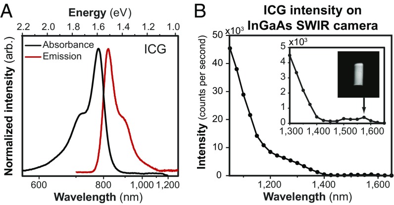
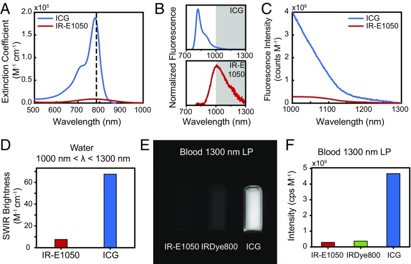
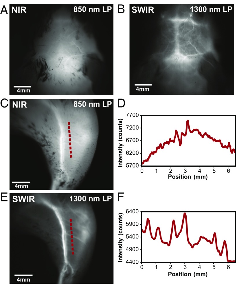
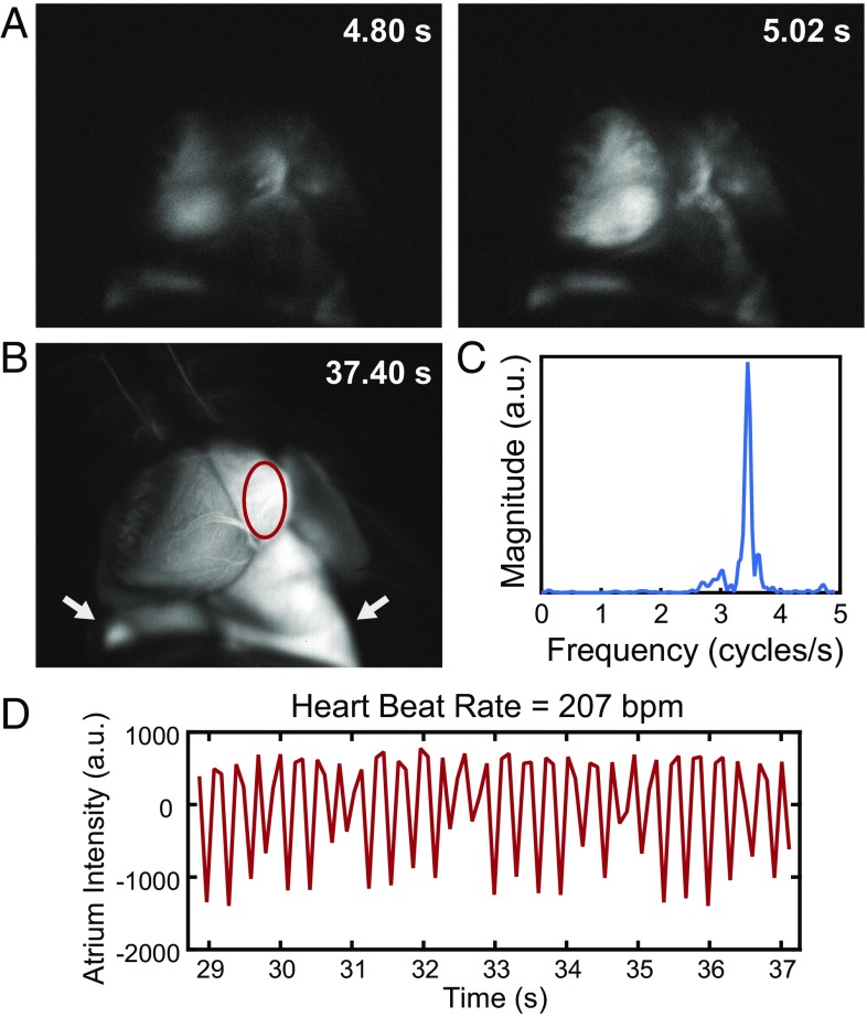
Fluorescence imaging is a method of real-time molecular tracking in vivo that has enabled many clinical technologies. Imaging in the shortwave IR (SWIR; 1,000-2,000 nm) promises higher contrast, sensitivity, and penetration depths compared with conventional visible and near-IR (NIR) fluorescence imaging. However, adoption of SWIR imaging in clinical settings has been limited, partially due to the absence of US Food and Drug Administration (FDA)-approved fluorophores with peak emission in the SWIR. Here, we show that commercially available NIR dyes, including the FDA-approved contrast agent indocyanine green (ICG), exhibit optical properties suitable for in vivo SWIR fluorescence imaging. Even though their emission spectra peak in the NIR, these dyes outperform commercial SWIR fluorophores and can be imaged in the SWIR, even beyond 1,500 nm. We show real-time fluorescence imaging using ICG at clinically relevant doses, including intravital microscopy, noninvasive imaging in blood and lymph vessels, and imaging of hepatobiliary clearance, and show increased contrast compared with NIR fluorescence imaging. Furthermore, we show tumor-targeted SWIR imaging with IRDye 800CW-labeled trastuzumab, an NIR dye being tested in multiple clinical trials. Our findings suggest that high-contrast SWIR fluorescence imaging can be implemented alongside existing imaging modalities by switching the detection of conventional NIR fluorescence systems from silicon-based NIR cameras to emerging indium gallium arsenide-based SWIR cameras. Using ICG in particular opens the possibility of translating SWIR fluorescence imaging to human clinical applications. Indeed, our findings suggest that emerging SWIR-fluorescent in vivo contrast agents should be benchmarked against the SWIR emission of ICG in blood.

Keywords: biomedical imaging; fluorescence imaging; indocyanine green; near infrared; shortwave infrared.


Conflict of interest statement

The authors declare no conflict of interest.

Figures

Fig. 1.
Fig. 1.
SWIR emission of the NIR dye ICG. (A) The full emission spectrum of ICG, measured on an NIR- and SWIR-sensitive InGaAs detector (red line), mirrors the absorption spectrum (black line) as predicted by the Franck–Condon principle. (B) The emission intensity of a 0.027 mg/mL aqueous ICG solution, detected in 20-nm spectral bands on an InGaAs camera and normalized by integration time, shows that emission is detectable up to at least 1,575 nm (Inset shows the vial). A lower intensity is observed between 1,400 and 1,500 nm due to the absorption band of water at those wavelengths.
Fig. 2.
Fig. 2.
Comparison of the optical properties of ICG with those of other NIR and SWIR dyes. (A) Normalized to identical molar concentrations, ICG absorbs much stronger at 785-nm excitation (dashed line) than IR-E1050, a commercially available SWIR dye marketed for in vivo SWIR imaging applications. (B) Thus, although the emission peak of ICG is significantly blue shifted compared with IR-E1050, (C) the measured emission intensity between 1,000 and 1,300 nm normalized to equimolar concentration is 8.7 times higher for ICG than for IR-E1050. (D) This finding was confirmed by calculating the SWIR brightness of ICG and IR-E1050 at wavelengths between 1,000 and 1,300 nm. Multiplying the independently measured fluorescence quantum yield, the maximum absorption cross-section, and the ratio of the number of photons emitted between 1,000 and 1,300 nm to the total number of emitted photons of both dyes shows that ICG is roughly nine times brighter than IR-E1050. (E) We further compared the fluorescence intensity of IR-E1050 (0.01 mg/mL) with that of IRDye 800CW PEG (0.01 mg/mL; not accounting for PEG shell of 25–60 kDa) and that of ICG (0.01 mg/mL) in bovine blood on an SWIR camera with a 1,300-nm long-pass (LP) filter. (F) Normalized to equimolar concentrations, the imaged intensity of the NIR dyes is greater than IR-E1050. SDs for signal intensity were found to be less than 5%. Fig. S2 and Movie S1 show comparisons in water and in vivo.
Fig. 3.
Fig. 3.
High contrast in vivo SWIR fluorescence imaging using ICG. (A) We noninvasively imaged the brain vasculature of a mouse using ICG contrast and find that the vessels are difficult to resolve through skin and skull using 850-nm long-pass (LP) NIR detection on a silicon camera. (B) Switching to 1,300-nm long-pass SWIR detection on an InGaAs camera greatly improves vessel contrast (Fig. S3 shows contrast quantification). (C) Similarly, only large hind-limb vessels are imaged with good contrast through the skin using NIR detection. (D) The intensity across a line of interest shows insufficient contrast to resolve smaller vessels from background signal. (E) Using 1,300-nm long-pass SWIR detection greatly improves image contrast and (F) resolution of vessels. All images were scaled to the maximum displayable intensities.
Fig. 4.
Fig. 4.
High temporal resolution shown through ICG SWIR fluorescence angiography. Intravital SWIR fluorescence angiography was performed in a mouse heart at 9.17 frames per second using ICG for contrast, diffuse 808-nm excitation, and a 1,300-nm long-pass emission filter on an InGaAs SWIR camera (Movie S2). (A) Temporal resolution was sufficiently high to resolve the heartbeat of the mouse. (B) By tracking a region of interest within the atrium of the heart (red circle; lungs are also pictured and indicated with white arrows) and (C) taking the Fourier transform of (D) the intensity fluctuations, the heart rate was determined to be 207 beats per minute for the anesthetized mouse. Fluorescence tracking details and assignment of anatomical structures are in Fig. S5.

References

    1. Sevick-Muraca EM. Translation of near-infrared fluorescence imaging technologies: Emerging clinical applications. Annu Rev Med. 2012;63:217–231. - PubMed
    1. Zhu B, Sevick-Muraca EM. A review of performance of near-infrared fluorescence imaging devices used in clinical studies. Br J Radiol. 2015;88:20140547. - PMC - PubMed
    1. Frangioni JV. In vivo near-infrared fluorescence imaging. Curr Opin Chem Biol. 2003;7:626–634. - PubMed
    1. Vahrmeijer AL, Hutteman M, van der Vorst JR, van de Velde CJH, Frangioni JV. Image-guided cancer surgery using near-infrared fluorescence. Nat Rev Clin Oncol. 2013;10:507–518. - PMC - PubMed
    1. Marshall MV, et al. Near-infrared fluorescence imaging in humans with indocyanine green: A review and update. Open Surg Oncol J. 2010;2:12–25. - PMC - PubMed

Publication types