. 2018 Apr:30:317-325.
doi: 10.1016/j.ebiom.2018.03.030. Epub 2018 Mar 28.

Sodium Butyrate Inhibits Inflammation and Maintains Epithelium Barrier Integrity in a TNBS-induced Inflammatory Bowel Disease Mice Model

Affiliations

Sodium Butyrate Inhibits Inflammation and Maintains Epithelium Barrier Integrity in a TNBS-induced Inflammatory Bowel Disease Mice Model

Guangxin Chen et al. EBioMedicine. 2018 Apr.

Abstract

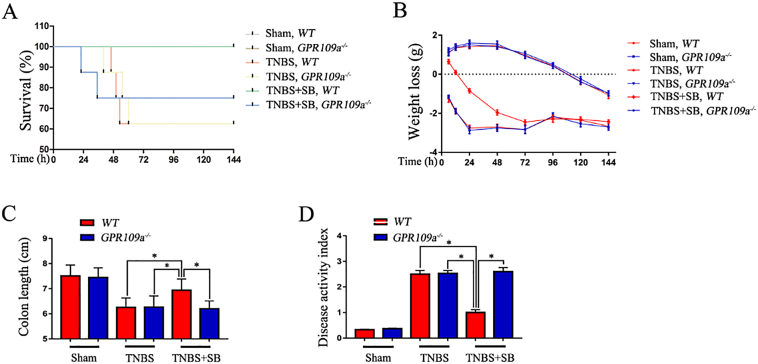
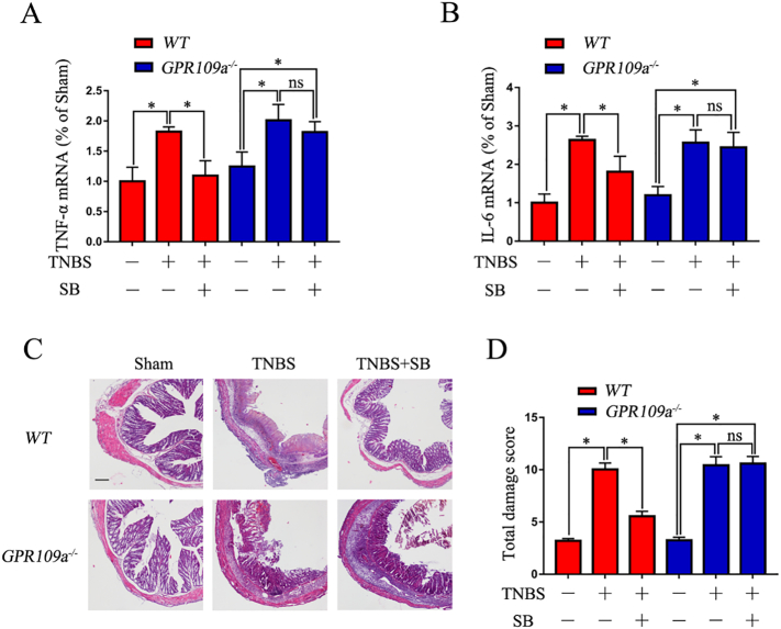
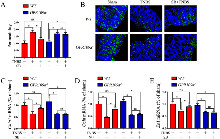
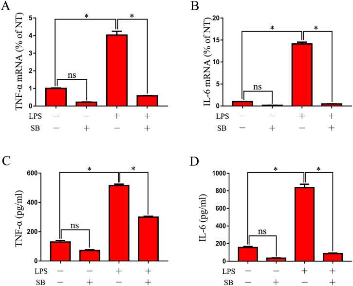
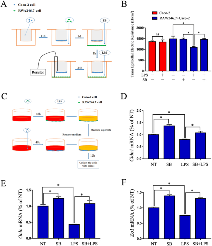
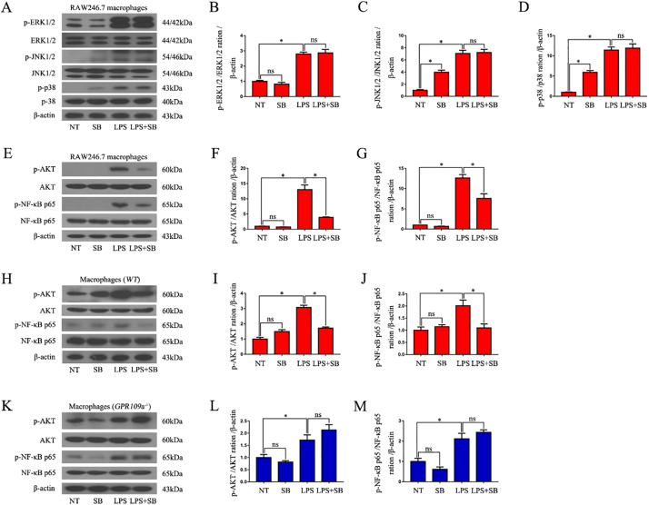
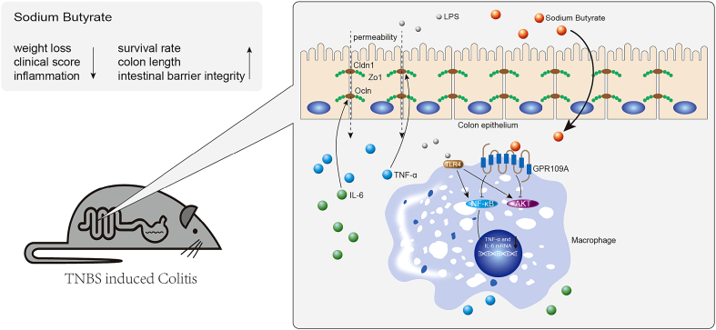
G Protein Coupled Receptor 109A (GPR109A), which belongs to the G protein coupled receptor family, can be activated by niacin, butyrate, and β-hydroxybutyric acid. Here, we assessed the anti-inflammatory activity of sodium butyrate (SB) on 2,4,6-trinitrobenzene sulfonic acid (TNBS)-induced colitis mice, an experimental model that resembles Crohn's disease, and explored the potential mechanism of SB in inflammatory bowel disease (IBD). In vivo, experimental GPR109a-/- and wild-type (WT) mice were administered SB (5g/L) in their drinking water for 6weeks. The mice were then administered TNBS via rectal perfusion to imitate colitis. In vitro, RAW246.7 macrophages, Caco-2 cells, and primary peritoneal macrophages were used to investigate the protective roles of SB on lipopolysaccharide (LPS)-induced inflammatory response and epithelium barrier dysfunction. In vivo, SB significantly ameliorated the inflammatory response and intestinal epithelium barrier dysfunction in TNBS-induced WT mice, but failed to provide a protective effect in TNBS-induced GPR109a-/- mice. In vitro, pre-treatment with SB dramatically inhibited the expression of TNF-α and IL-6 in LPS-induced RAW246.7 macrophages. SB inhibited the LPS-induced phosphorylation of the NF-κB p65 and AKT signaling pathways, but failed to inhibit the phosphorylation of the MAPK signaling pathway. Our data indicated that SB ameliorated the TNBS-induced inflammatory response and intestinal epithelium barrier dysfunction through activating GPR109A and inhibiting the AKT and NF-κB p65 signaling pathways. These findings therefore extend the understanding of GPR109A receptor function and provide a new theoretical basis for treatment of IBD.

Keywords: Epithelium barrier; GPR109A; IBD; Inflammation; SB; TNBS.


Figures

Fig. 1
Fig. 1
SB ameliorates survival, weight loss, colon length, and disease activity index in the TNBS-induced mouse IBD model. WT and GPR109a−/− mice were pre-treated with SB for 6 weeks and then treated with TNBS for 6 days. (A) Survival of TNBS- and SB-treated WT and GPR109a−/−mice (n = 14). (B) Weight loss (the previous day's body weight – the day's body weight) in TNBS- and SB-treated WT and GPR109a−/−mice (n = 8–12). (C) Colon length in TNBS- and SB-treated WT and GPR109a−/− mice (n = 8–12). (D) Disease activity index of TNBS- and SB-treated WT and GPR109a−/−mice (n = 8–12).
Fig. 2
Fig. 2
SB suppresses the inflammation in TNBS-induced mice. (A-B) Expression of the pro-inflammatory cytokines TNF-α and IL-6 in colon tissue homogenate (n = 4). (C) H&E staining of colon tissue from TNBS- and SB-treated WT and GPR109a−/−mice, magnification shown is 4 ×, and the scale bar represents 100 μm (n = 4). (D) Total damage score of TNBS- and SB-treated WT and GPR109a−/− mice (n = 8–12).
Fig. 3
Fig. 3
SB reduces the permeability and maintains proper tight junctions and mucin-2 in TNBS-induced mice. All mice were pre-treated with SB for 6 weeks, and then treated with TNBS for 2 days. (A) Relative permeability of TNBS and SB treatment in WT and GPR109a−/− mice. After treating with SB and TNBS, the mice were starved for 4 h and then gavaged with FITC-dextran (0.6 mg/g body weight at a concentration of 125 mg/mL). Serum from blood collected after 4 h was used to measure the permeability (n = 4). (B) Immunofluorescence staining of Muc2 in the colon; magnification shown is 10 ×, and the scale bar represents 300 μm (n = 4). (C-E) Expression of the tight junction encoding genes Cldn1, Ocln, and Zo1 in the colon tissue homogenate of TNBS- and SB-treated WT and GPR109a−/− mice (n = 4).
Fig. 4
Fig. 4
SB suppresses pro-inflammatory cytokine expression in LPS-induced macrophages. (A-B) Expression of TNF-α and IL-6 in SB- and LPS-treated RAW246.7 cells. RAW246.7 macrophages were stimulated with LPS for 6 h after pre-treating with SB for 1 h, and then the cells were collected with TRIzol. Gene expression of TNF-α and IL-6 was detected by qRT-PCR (n = 3). (C-D) Protein levels of TNF-α and IL-6 in the medium supernatant of SB- and LPS-treated RAW246.7 macrophages. RAW246.7 macrophages were pre-treated with SB for 1 h, and then stimulated with LPS for 12 h. The medium supernatant was collected and used for measuring the protein levels of TNF-α and IL-6 by ELISA (n = 3).
Fig. 5
Fig. 5
SB maintains a proper intestinal epithelium barrier by inhibiting the inflammatory response of macrophages. (A) Schematic diagram of the testing process of TEER. (B) TEER of various treatments of the Caco-2 monolayer (n = 3). (C) Schematic diagram of Caco-2 cell and RAW246.7 macrophage co-culture. (D-E) Expression of the tight junction genes Cldn1, Ocln, and Zo1 of Caco-2 cells (n = 3).
Fig. 6
Fig. 6
SB suppresses LPS-induced phosphorylation of the AKT and NF-κB p65 signaling pathways in macrophages. (A-G) Western blot analysis of the phosphorylation of the p38, ERK p-38, JNK1/2, ERK1/2, AKT, and NF-κB p65 signaling pathways in LPS-induced RAW246.7 macrophages (n = 3). (H-J) Western blot analysis of the phosphorylation of the AKT and NF-κB p65 signaling pathways in LPS-induced WT mouse primary peritoneal macrophages (n = 3). (K-M) Western blot analysis of the phosphorylation of the AKT and NF-κB p65 signaling pathways in LPS-induced GPR109a−/−mouse primary peritoneal macrophages (n = 3).
Fig. 7
Fig. 7
Mechanism of sodium butyrate on inhibiting inflammation and maintaining epithelium barrier integrity in the TNBS-induced colitis model.

References

    1. Artis D., Grencis R.K. The intestinal epithelium: sensors to effectors in nematode infection. Mucosal Immunol. 2008;1:252–264. - PubMed
    1. Baumgart D.C., Sandborn W.J. Crohn's disease. Lancet. 2012;380:1590–1605. - PubMed
    1. Beaugerie L., Seksik P., Nion-Larmurier I., Gendre J.P., Cosnes J. Predictors of Crohn's disease. Gastroenterology. 2006;130:650–656. - PubMed
    1. Cantoni S., Galletti M., Zambelli F., Valente S., Ponti F., Tassinari R., Pasquinelli G., Galie N., Ventura C. Sodium butyrate inhibits platelet-derived growth factor-induced proliferation and migration in pulmonary artery smooth muscle cells through Akt inhibition. FEBS J. 2013;280:2042–2055. - PubMed
    1. Chen J., Li Y., Tian Y., Huang C., Li D., Zhong Q., Ma X. Interaction between microbes and host intestinal health: modulation by dietary nutrients and gut-brain-endocrine-immune axis. Curr. Protein Pept. Sci. 2015;16:592–603. - PubMed

MeSH terms