

RNA Binding Proteins in Intestinal Epithelial Biology and Colorectal Cancer
- PMID: 29627433
- PMCID: PMC5927824
- DOI: 10.1016/j.molmed.2018.03.008
RNA Binding Proteins in Intestinal Epithelial Biology and Colorectal Cancer
Abstract
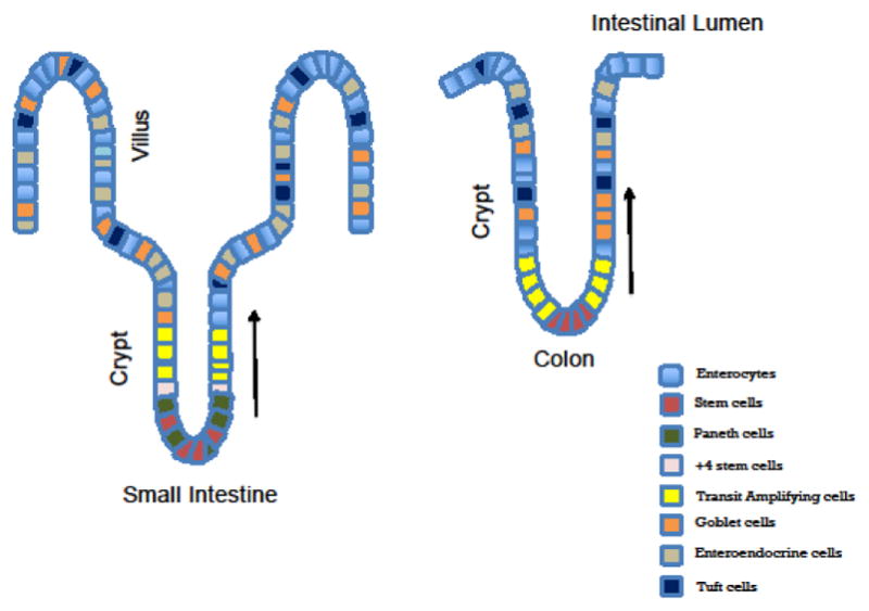
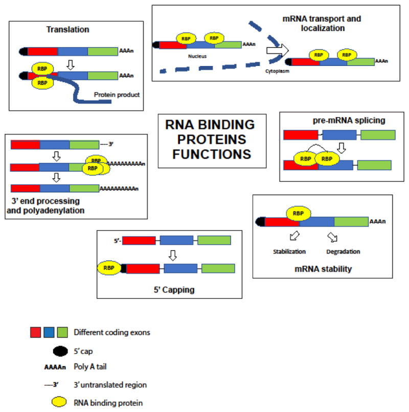
The intestinal epithelium is highly proliferative and consists of crypt invaginations that house stem cells and villus projections with differentiated cells. There exists a dynamic equilibrium between proliferation, migration, differentiation, and senescence that is regulated by several factors. Among these are RNA binding proteins (RBPs) that bind their targets in a both context dependent and independent manner. RBP-RNA complexes act as rheostats by regulating expression of RNAs both co- and post-transcriptionally. This is important, especially in response to intestinal injury, to fuel regeneration. The manner in which these RBPs function in the intestine and their interactions with other pivotal pathways in colorectal cancer may provide a framework for new insights and potential therapeutic applications.
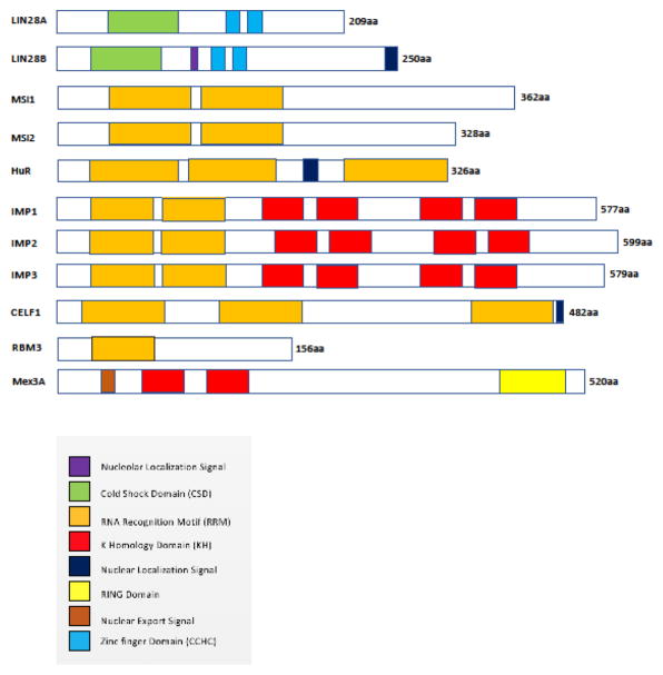
Keywords: CUGBP Elav-Like Family Member 1 (CELF1); LIN28; MEX3A; Musashi (MSI); RNA binding protein 3 (RBM3) and Hu-Antigen R (HUR); RNA binding proteins; colorectal cancer; insulin-like growth factor 2 mRNA binding proteins (IGF2BP/IMP); intestinal stem cells.
Copyright © 2018 Elsevier Ltd. All rights reserved.
Figures
References
-
- van der Flier LG, Clevers H. Stem cells, self-renewal, and differentiation in the intestinal epithelium. Annu Rev Physiol. 2009;71:241–60. - PubMed
-
- Gregorieff A, et al. Expression pattern of Wnt signaling components in the adult intestine. Gastroenterology. 2005;129(2):626–38. - PubMed
-
- van der Flier LG, et al. Transcription factor achaete scute-like 2 controls intestinal stem cell fate. Cell. 2009;136(5):903–12. - PubMed
Publication types
MeSH terms
Substances
Grants and funding
LinkOut - more resources
Full Text Sources
Other Literature Sources
Medical
Research Materials
Miscellaneous
