

Snail-borne parasitic diseases: an update on global epidemiological distribution, transmission interruption and control methods
- PMID: 29628017
- PMCID: PMC5890347
- DOI: 10.1186/s40249-018-0414-7
Snail-borne parasitic diseases: an update on global epidemiological distribution, transmission interruption and control methods
Abstract
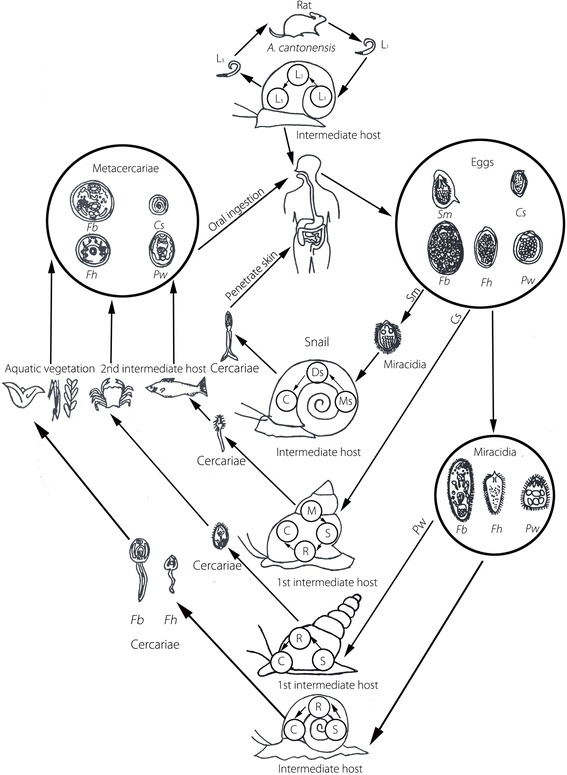
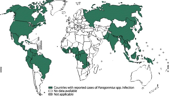
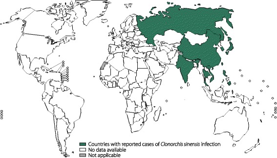
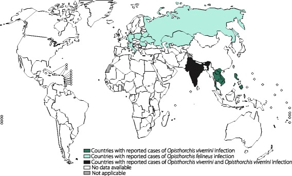
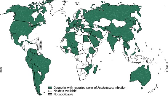
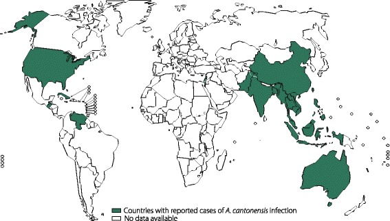
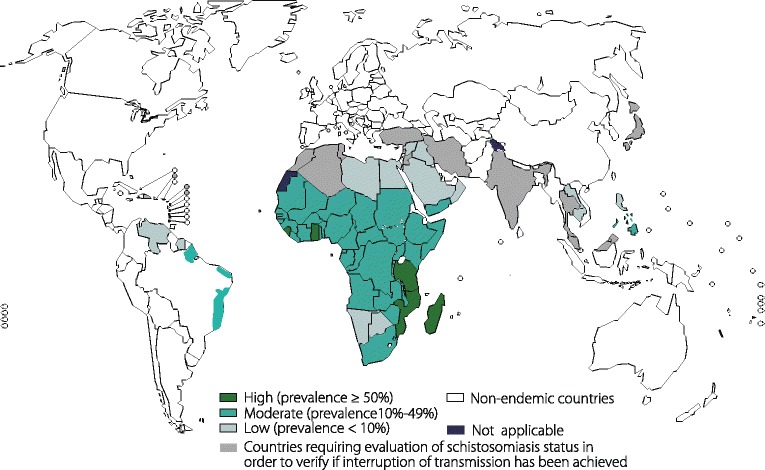
Background: Snail-borne parasitic diseases, such as angiostrongyliasis, clonorchiasis, fascioliasis, fasciolopsiasis, opisthorchiasis, paragonimiasis and schistosomiasis, pose risks to human health and cause major socioeconomic problems in many tropical and sub-tropical countries. In this review we summarize the core roles of snails in the life cycles of the parasites they host, their clinical manifestations and disease distributions, as well as snail control methods.
Main body: Snails have four roles in the life cycles of the parasites they host: as an intermediate host infected by the first-stage larvae, as the only intermediate host infected by miracidia, as the first intermediate host that ingests the parasite eggs are ingested, and as the first intermediate host penetrated by miracidia with or without the second intermediate host being an aquatic animal. Snail-borne parasitic diseases target many organs, such as the lungs, liver, biliary tract, intestines, brain and kidneys, leading to overactive immune responses, cancers, organ failure, infertility and even death. Developing countries in Africa, Asia and Latin America have the highest incidences of these diseases, while some endemic parasites have developed into worldwide epidemics through the global spread of snails. Physical, chemical and biological methods have been introduced to control the host snail populations to prevent disease.
Conclusions: In this review, we summarize the roles of snails in the life cycles of the parasites they host, the worldwide distribution of parasite-transmitting snails, the epidemiology and pathogenesis of snail-transmitted parasitic diseases, and the existing snail control measures, which will contribute to further understanding the snail-parasite relationship and new strategies for controlling snail-borne parasitic diseases.
Keywords: Epidemiology; Pathogenesis; Snail control; Snail-borne parasitic diseases.
Conflict of interest statement
Ethics approval and consent to participate
Not applicable.
Consent for publication
Not applicable.
Competing interests
The authors declare that they have no competing interests.
Figures

References
Publication types
MeSH terms
Grants and funding
LinkOut - more resources
Full Text Sources
Other Literature Sources
Medical
Research Materials
