. 2018 May 3;173(4):958-971.e17.
doi: 10.1016/j.cell.2018.03.025. Epub 2018 Apr 5.

Stress Granule Assembly Disrupts Nucleocytoplasmic Transport

Affiliations

Stress Granule Assembly Disrupts Nucleocytoplasmic Transport

Ke Zhang et al. Cell. .

Abstract

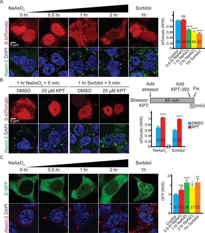
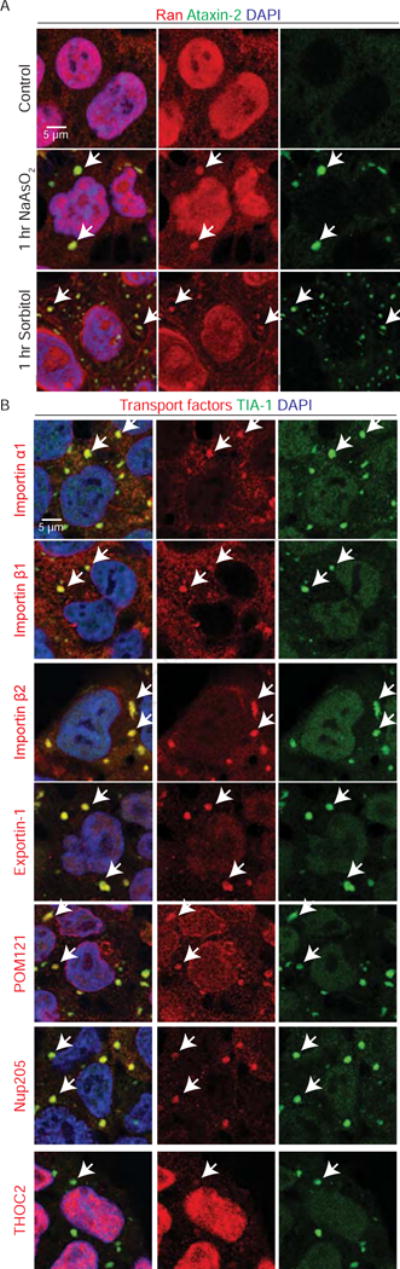
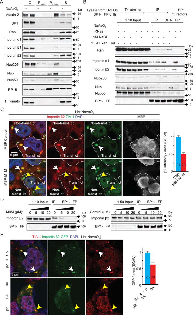
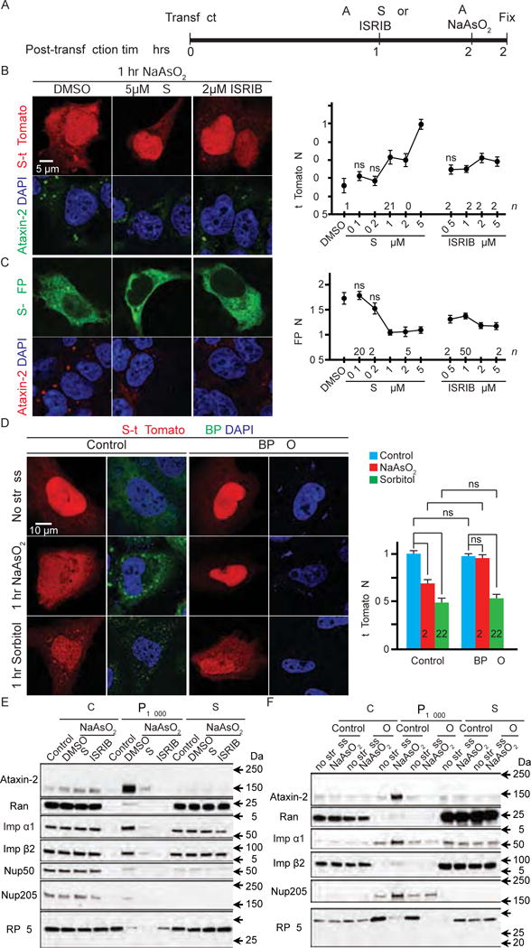
Defects in nucleocytoplasmic transport have been identified as a key pathogenic event in amyotrophic lateral sclerosis (ALS) and frontotemporal dementia (FTD) mediated by a GGGGCC hexanucleotide repeat expansion in C9ORF72, the most common genetic cause of ALS/FTD. Furthermore, nucleocytoplasmic transport disruption has also been implicated in other neurodegenerative diseases with protein aggregation, suggesting a shared mechanism by which protein stress disrupts nucleocytoplasmic transport. Here, we show that cellular stress disrupts nucleocytoplasmic transport by localizing critical nucleocytoplasmic transport factors into stress granules, RNA/protein complexes that play a crucial role in ALS pathogenesis. Importantly, inhibiting stress granule assembly, such as by knocking down Ataxin-2, suppresses nucleocytoplasmic transport defects as well as neurodegeneration in C9ORF72-mediated ALS/FTD. Our findings identify a link between stress granule assembly and nucleocytoplasmic transport, two fundamental cellular processes implicated in the pathogenesis of C9ORF72-mediated ALS/FTD and other neurodegenerative diseases.

Keywords: ALS; C9ORF72; nucleocytoplasmic transport; stress granule.


Conflict of interest statement

Declaration of interests: J.P.T. received support from Inception Sciences. The remaining authors declare no competing interests.

Figures

Figure 1
Figure 1. Stressors disrupt nucleocytoplasmic transport
(A) HEK293T cells expressing S-tdTomato (red) stained with Ataxin-2 (green) and DAPI (blue). (B) Stressed HEK293T cells expressing S-tdTomato (red) treated with KPT-350 or DMSO and stained with Ataxin-2 (green) and DAPI (blue). (C) HEK293T cells expressing S-GFP stained with Ataxin-2 (red), GFP (green), and DAPI (blue). N: nuclear; W: whole cell. n numbers in graph. ns: not significant *: p<0.05; **: p<0.01; ****: p<0.0001. Data are represented as mean ± SEM.
Figure 2
Figure 2. Nucleocytoplasmic transport factors are localized to stress granules
(A) Untreated (top row), arsenite- (middle row) or sorbitol- (bottom row) treated HEK293T cells stained with Ran (red), Ataxin-2 (green) and DAPI (blue). Arrows indicate co-localization. (B) Arsenite-treated HEK293T cells stained with transport factors (red), TIA-1 (green) and DAPI (blue). Arrows indicate co-localization.
Figure 3
Figure 3. Nucleocytoplasmic transport factors are constituents of stress granules
(A) Subcellular fractionation of HEK293T cells. P1000: pellet from 1,000 g; P18000: pellet from 18,000 g; S: supernatant after 18,000 g. (B) Co-IP of nucleocytoplasmic transport factors with G3BP1-GFP from U-2 OS cells expressing G3BP1-GFP. (C) HEK293T cells expressing MBP (control, top) or MBP-tagged M9M (bottom) were stained with Importin β2 (red), TIA-1 (green), DAPI (blue), and MBP (white). Dashed lines separate transfected versus non-transfected cells. White arrowheads indicate co-localization. Yellow arrowheads indicate TIA-1-positive puncta without Importin β2 co-localization. (D) Co-IP of Importin β2 and G3BP1-GFP with chemically synthesized M9M or control peptide. (E) HEK293T cells expressing GFP-tagged wild type (top) or mutant (bottom) Importin β2 (green) stained with TIA-1 (red) and DAPI (blue). Arrowheads indicate co-localization. W: whole cell, n numbers in graph. ****: p<0.0001. Data are represented as mean ± SEM.
Figure 4
Figure 4. Stress granules mediate the nucleocytoplasmic transport defects caused by arsenite
(A) Experimental design. (B and C) HEK293T cells expressing S-tdTomato (red) (B) or S-GFP (green) (C) were treated with arsenite and GSK or ISRIB and stained with Ataxin-2 (green in B and red in C) and DAPI (blue). (D) Control (left two columns) or G3BP1/2 double knockout (G3BP KO) U-2 OS cells (right two columns) expressing S-tdTomato (red) stained with G3BP (green) and (DAPI). N: nuclear; W: whole cell. (E and F) Subcellular fractionation of control and arsenite-treated HEK293T cells with or without GSK or ISRIB pre-treatment (E) or wild-type control or G3BP KO (KO) U-2 OS cells (F). WCL: whole cell lysate; P18000: pellet from 18,000 g; S: supernatant after 18,000 g. n numbers in graph. ns: not significant; *: p<0.05; **: p<0.01; ****: p<0.0001. Data are represented as mean ± SEM.
Figure 5
Figure 5. Dipeptide repeat proteins and cytoplasmic TDP-43 cause nucleocytoplasmic transport defects
(A) HEK293T cells co-expressing S-tdTomato (red) with GFP (left column), GFP-tagged 50 repeats of poly-GR (column 2) or poly-PR (column 3), or cytoplasmic TDP-43 (TDP(cyto)) (right column) stained with DAPI (blue). N: nuclear; W: whole cell. (B) HEK293T cells expressing GFP (top row), GFP-tagged (GR)50 (row 2) or (PR)50 (row 3), or TDP(cyto) (bottom row) were stained with Ran (red), Ataxin-2 (green) and DAPI (blue). GFP expression shown on right (white). Arrowheads indicate co-localization. Number of cells measured (n) for each condition indicated in graph. *: p<0.05; ****: p<0.0001. Data are represented as mean ± SEM.
Figure 6
Figure 6. Stress granules contribute to nucleocytoplasmic transport defects caused by poly-GR, poly-PR or cytoplasmic TDP-43
(A) HEK293T cells co-expressing S-tdTomato (red) and GFP-tagged (GR)50, (PR)50, or cytoplasmic TDP-43 (TDP(cyto)) (green) treated with DMSO, GSK or ISRIB and stained with DAPI (blue). Quantification in B. (C) Wild type control or G3BP1/2 double knockout (G3BP KO) U-2 OS cells co-expressing S-tdTomato (red) and GFP-tagged (GR)50, (PR)50, or TDP(cyto) (green) stained with DAPI (blue). Quantification in D. N: nuclear; W: whole cell. n numbers in graph. *: p<0.05; **: p<0.01; ***: p<0.001; ****: p<0.0001. Data are represented as mean ± SEM.
Figure 7
Figure 7. GSK, ISRIB, and Ataxin-2 ASO suppress nucleocytoplasmic transport defects and neurodegeneration in C9-ALS models
(A) C9-ALS iPSNs treated with DMSO, GSK or ISRIB were stained with Ran (red), MAP2 (green), and DAPI (blue). (B) Control or C9-ALS iPSNs treated with scrambled or Ataxin-2 ASOs were stained with Ran (red), MAP2 (green), and DAPI (blue). Bottom right: ASO-treated C9-ALS iPSNs immunoblotted for Ataxin-2 and β-Actin. (C) Fly salivary glands stained with GFP and DAPI. N: nuclear; W: whole cell. (D) Fly eye degeneration. (E) Flight assay. n numbers in the graph. ns: not significant; *: p<0.05; **: p<0.01; ***: p<0.001; ****: p<0.0001. Data are represented as mean ± SEM.

Similar articles

Cited by

References

    1. Anderson P, Kedersha N. Stress granules: the Tao of RNA triage. Trends Biochem Sci. 2008;33:141–150. - PubMed
    1. Apicco DJ, Ash PEA, Maziuk B, LeBlang C, Medalla M, Al AA, Ferragud A, Botelho E, Ballance HI, Dhawan U, et al. Reducing the RNA binding protein TIA1 protects against tau-mediated neurodegeneration in vivo. Nat Neurosci. 2018;21:72–80. - PMC - PubMed
    1. Ash PE, Bieniek KF, Gendron TF, Caulfield T, Lin WL, DeJesus-Hernandez M, van Blitterswijk MM, Jansen-West K, Paul JW, III, Rademakers R, et al. Unconventional translation of C9ORF72 GGGGCC expansion generates insoluble polypeptides specific to c9FTD/ALS. Neuron. 2013;77:639–646. - PMC - PubMed
    1. Axten JM, Medina JR, Feng Y, Shu A, Romeril SP, Grant SW, Li WH, Heerding DA, Minthorn E, Mencken T, et al. Discovery of 7-methyl-5-(1-{[3-(trifluoromethyl)phenyl]acetyl}-2,3-dihydro-1H-indol-5-yl)-7H-p yrrolo[2,3-d]pyrimidin-4-amine (GSK2606414), a potent and selective first-in-class inhibitor of protein kinase R (PKR)-like endoplasmic reticulum kinase (PERK) J Med Chem. 2012;55:7193–7207. - PubMed
    1. Banani SF, Lee HO, Hyman AA, Rosen MK. Biomolecular condensates: organizers of cellular biochemistry. Nat Rev Mol Cell Biol. 2017;18:285–298. - PMC - PubMed

Publication types

MeSH terms

Supplementary concepts