. 2018 Jun 8;293(23):8934-8946.
doi: 10.1074/jbc.M117.817031. Epub 2018 Apr 8.

Structural analyses of Arabidopsis thaliana legumain γ reveal differential recognition and processing of proteolysis and ligation substrates

Affiliations

Structural analyses of Arabidopsis thaliana legumain γ reveal differential recognition and processing of proteolysis and ligation substrates

Florian B Zauner et al. J Biol Chem. .

Abstract

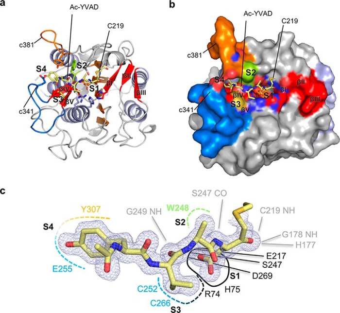
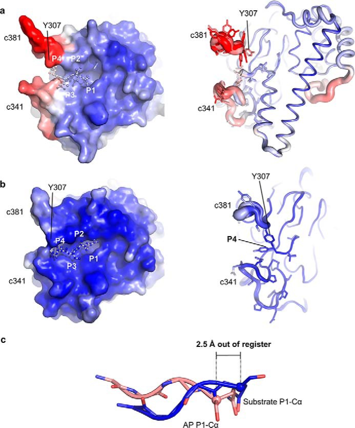
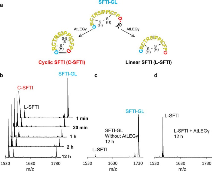
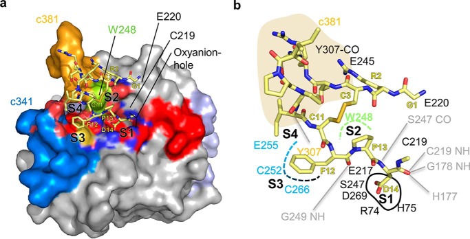
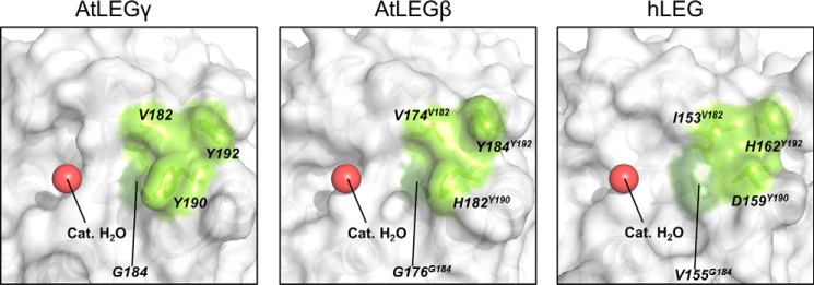
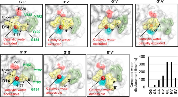
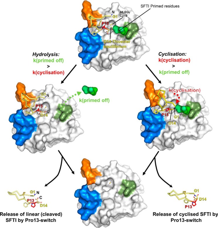
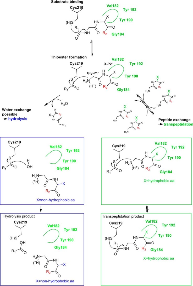
Legumain is a dual-function protease-peptide ligase whose activities are of great interest to researchers studying plant physiology and to biotechnological applications. However, the molecular mechanisms determining the specificities for proteolysis and ligation are unclear because structural information on the substrate recognition by a fully activated plant legumain is unavailable. Here, we present the X-ray structure of Arabidopsis thaliana legumain isoform γ (AtLEGγ) in complex with the covalent peptidic Ac-YVAD chloromethyl ketone (CMK) inhibitor targeting the catalytic cysteine. Mapping of the specificity pockets preceding the substrate-cleavage site explained the known substrate preference. The comparison of inhibited and free AtLEGγ structures disclosed a substrate-induced disorder-order transition with synergistic rearrangements in the substrate-recognition sites. Docking and in vitro studies with an AtLEGγ ligase substrate, sunflower trypsin inhibitor (SFTI), revealed a canonical, protease substrate-like binding to the active site-binding pockets preceding and following the cleavage site. We found the interaction of the second residue after the scissile bond, P2'-S2', to be critical for deciding on proteolysis versus cyclization. cis-trans-Isomerization of the cyclic peptide product triggered its release from the AtLEGγ active site and prevented inadvertent cleavage. The presented integrative mechanisms of proteolysis and ligation (transpeptidation) explain the interdependence of legumain and its preferred substrates and provide a rational framework for engineering optimized proteases, ligases, and substrates.

Keywords: chemical biology; computational biology; crystal structure; cysteine protease; pH regulation; peptide biosynthesis; plant biochemistry; structural biology; transpeptidation; water displacement model.


Conflict of interest statement

The authors declare that they have no conflicts of interest with the contents of this article

Figures

Figure 1.
Figure 1.
Interaction topology of the peptidic Ac-YVAD-CMK inhibitor with fully activated AtLEGγ (AEP). The active form of AtLEGγ is displayed in standard orientation, i.e. with substrate running from left to right. The specificity loops c341 and c381 are colored in blue or orange, respectively. a, complex structure of the covalently bound Ac-YVAD-CMK-based inhibitor in a cartoon model. b, surface representation of a showing how the specificity loops embrace the substrate. c, mapping of the substrate recognition sites. The ligand and its electron density (2FoFc map, 1σ contouring) are shown in blue. The pockets and its main interactors are indicated with color code: black, S1; green, S2; blue, S3; orange, S4. The backbone interactions are shown in dashed lines.
Figure 2.
Figure 2.
Substrate recognition in the fully activated AtLEGγ (AEP) form differs from the activation peptide (AP) binding in the zymogenic two-chain state with respect to both structure and dynamics. Surface (left) and cartoon representations are used to color code the conformational variabilities by their crystallographic temperature factors. The color spectrum from blue to red represents low to high conformational variability, i.e. rigid (blue) to flexible (red). a, note the high temperature factors and multiple conformations of the specificity loops c341 and c381 in the zymogenic two-chain state. b, the fully activated catalytic domain shows well-ordered specificity loops. c, structural superposition of the activation peptide of the zymogenic two-chain state (AP) in orange and the substrate Ac-YVAD in blue. Note the shift in register of around 2.5 Å at the P1 Cα positions.
Figure 3.
Figure 3.
AtLEGγ efficiently cyclizes SFTI-GL but not cleaved SFTI. a, basic experiment design at pH 6.5. b, time course cyclization of SFTI-GL (reduced and oxidized) by AtLEGγ and control experiments. No cyclic SFTI was observed when the SFTI-GL was incubated without AtLEGγ. c or d, when L-SFTI was incubated with AtLEGγ.
Figure 4.
Figure 4.
Docking reveals canonical binding mode of SFTI. a, top view on the docked complex. b, interaction map of docked SFTI. Gray lines indicate backbone interactions.
Figure 5.
Figure 5.
S2′ pockets for different legumain isoforms and position of the catalytic water. Different legumain isoforms are displayed in surface representation. Bright and dark green are the wall- and bottom-forming residues of the different legumain S2′ pockets, respectively. A red sphere indicates the position of the catalytic water. The PDB ID of human legumain (hLEG) is 4AWA. The structure of AtLEGβ has been built as an homology model using AtLEGγ as a starting model. The homology model of the catalytic domain of AtLEGβ was built using the software Phyre2 (54).
Figure 6.
Figure 6.
S2′ pocket of plant legumains has high affinity for hydrophobic P2′ residues, critical for water displacement. All structures are results of 330-ns molecular dynamics simulations with identical starting structures, despite the indicated mutation of the primed residues. The different primed tails are labeled above each structure. Yellow, representation of the still bound product, which is thioester-bound to Cys219 via Asp14; the released primed peptides at the end of the simulation are also indicated. Cyan, catalytic histidine. Red, Glu220. Dark green, S2′ pocket. The putative catalytic water is highlighted. The individual computed water exclusion times are also shown.
Figure 7.
Figure 7.
Water exclusion model of SFTI cyclization and proteolysis. Top, SFTI-GL binds canonically (like a substrate) to AtLEGγ. The primed GL tail occupies the S2′ pocket. The N terminus (Gly1) is positioned close to the catalytic cysteine. The attack of the catalytic cysteine creates the acyl-enzyme intermediate, which can be stabilized by the GL-tail shielding it from water. Left, cleavage is achieved when the primed tail of SFTI cannot productively interact with the S2′ pocket, which leads to hydrolysis of the thioester, resulting in linear SFTI (L-SFTI). Right, the N terminus of SFTI can attack the thioester and cyclic SFTI (C-SFTI) is produced. The Pro13 cis-trans-isomerization triggers the efficient off-dissociation. Bottom, the products of the hydrolysis and the cyclization reaction (shown at the bottom left and bottom right, respectively) are released from enzyme AtLEGγ (bottom center).
Figure 8.
Figure 8.
Mechanistic scheme of transpeptidation and hydrolysis. Green rectangles show transpeptidation reactions induced by a hydrophobic residue in P2′. The hydrolysis reaction pathway is indicated by cyan rectangles, induced by a nonhydrophobic residue in P2′. Starting point: green dashed rectangle. A substrate (R1 + R2) binds to the active site of AtLEGγ. The attack of Cys219 creates a thioester and free N terminus at the primed site. Left pathway, if the initially bound substrate (green dashed line) carries a nonhydrophobic residue in P2′, the primed product can dissociate after the formation of the thioester and water will exchange. Consequently, this results in hydrolysis of the thioester and the release of the hydrolysis product. Right pathway, if P2′ is hydrophobic, the primed site peptide stays bound and prevents the exchange of catalytic water, resulting in an equilibrium between thioester and peptide bond. In the presence of a suitable transpeptidation substrate (R3), an exchange between the initially bound primed product and the transpeptidation peptide can happen. This results in a new equilibrium between thioester and peptide bond, forming the transpeptidation product. The varying protonation state of the released primed N terminus is indicated. Only a deprotonated N terminus is able to attack the thioester, not a protonated one. This relationship explains the pH dependence of transpeptidation, which is more efficient at neutral pH than acidic pH.

References

    1. Conlan B. F., Gillon A. D., Craik D. J., and Anderson M. A. (2010) Circular proteins and mechanisms of cyclization. Biopolymers 94, 573–583 10.1002/bip.21422 - DOI - PubMed
    1. Craik D. J., and Malik U. (2013) Cyclotide biosynthesis. Curr. Opin. Chem. Biol. 17, 546–554 10.1016/j.cbpa.2013.05.033 - DOI - PubMed
    1. Dall E., and Brandstetter H. (2016) Structure and function of legumain in health and disease. Biochimie (Paris) 122, 126–150 10.1016/j.biochi.2015.09.022 - DOI - PubMed
    1. Gillon A. D., Saska I., Jennings C. V., Guarino R. F., Craik D. J., and Anderson M. A. (2008) Biosynthesis of circular proteins in plants. Plant J. 53, 505–515 - PubMed
    1. Müntz K., and Shutov A. D. (2002) Legumains and their functions in plants. Trends Plant Sci. 7, 340–344 10.1016/S1360-1385(02)02298-7 - DOI - PubMed

Publication types