

Review
doi: 10.1055/s-0038-1636516.
Epub 2018 Apr 5.
Updates on MR-Guided Focused Ultrasound for Symptomatic Uterine Fibroids
Affiliations
- PMID: 29628611
- PMCID: PMC5886770
- DOI: 10.1055/s-0038-1636516
Item in Clipboard
Review
Updates on MR-Guided Focused Ultrasound for Symptomatic Uterine Fibroids
Semin Intervent Radiol.
2018 Mar.
Abstract
Magnetic-resonance-guided focused ultrasound (MRgFUS), also called high-intensity focused ultrasound (HIFU) is an effective, noninvasive uterine-preserving treatment for symptomatic uterine fibroids. As the use of this therapeutic modality is not yet widespread, it may remain unfamiliar to many interventional radiologists. The purpose of this review is to discuss MRgFUS, including technology, patient selection, technique, outcomes, complications, and recent data on fertility and comparative effectiveness.
Keywords: High-intensity focused ultrasound; fibroids; interventional radiology; menorrhagia.
Figures
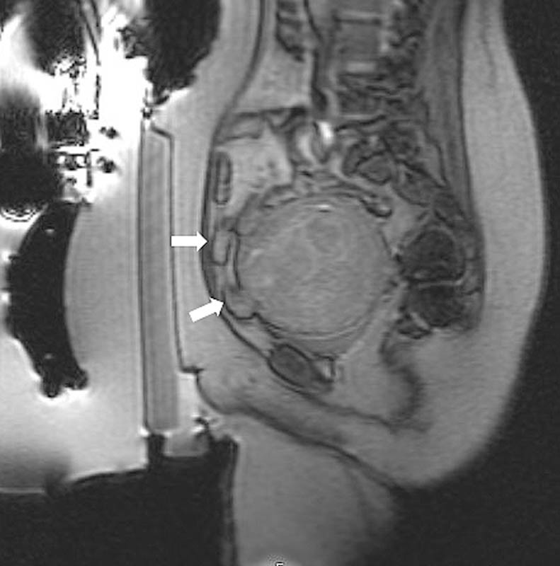
Sagittal GRE image demonstrates loops of intestine (white arrows) anterior to the fibroid, in the intended treatment path.
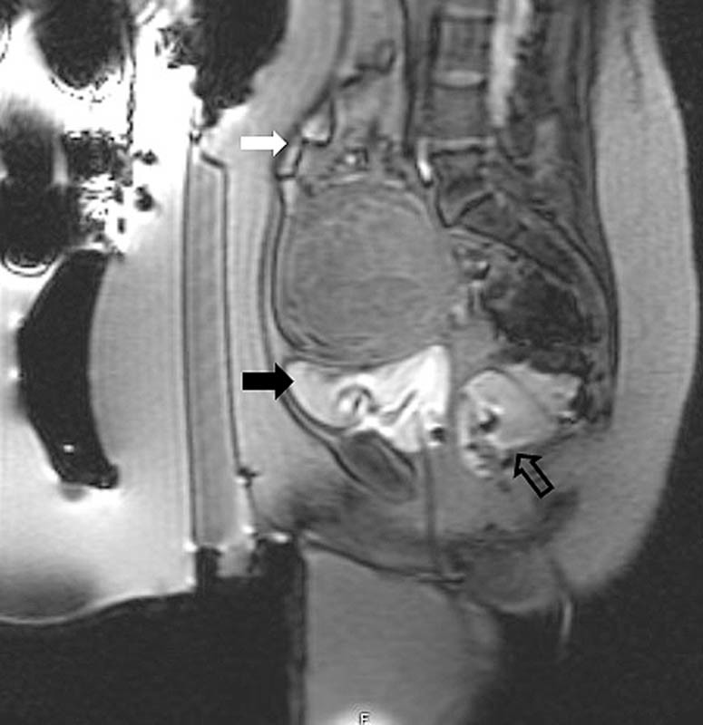
Following filling of the bladder (black arrow) and rectum (open arrow), the intestine (white arrow) has been displaced superiorly and out of the treatment path.
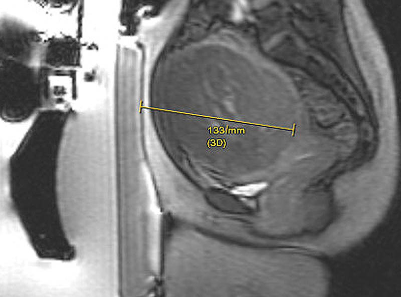
Sagittal GRE image demonstrates the distance from the skin to the posterior aspect of the fibroid to be 13.3 cm, which is outside the 12 cm range recommended for optimal therapy.
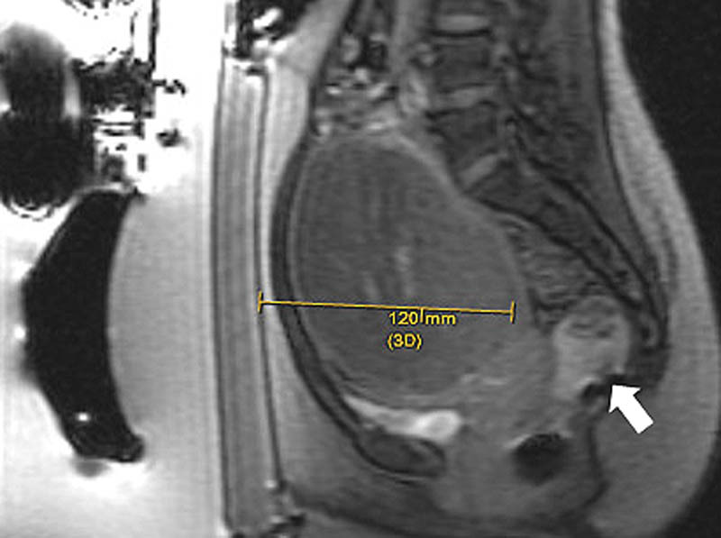
Following rectal fill (arrow), the uterus and the fibroid are displaced anteriorly, within the recommended 12 cm range.
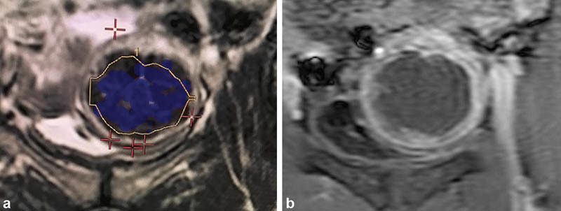
The blue overlay (
a
) corresponds to the nonenhancing portion of the fibroid (
b
).
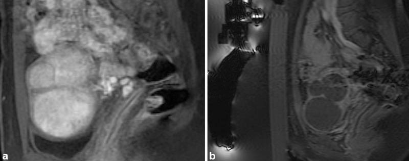
Sagittal T1-weighted postcontrast fast-saturated images. (
a
) Multiple enhancing fibroids are present in the uterus. (
b
) Following magnetic-resonance–guided focused ultrasound, none of the fibroids are enhancing resulting in a high nonperfused volume.
Similar articles
-
Clinical and Technical Aspects of MR-Guided High Intensity Focused Ultrasound for Treatment of Symptomatic Uterine Fibroids.Semin Intervent Radiol. 2013 Dec;30(4):347-53. doi: 10.1055/s-0033-1359728. Semin Intervent Radiol. 2013. PMID: 24436561 Free PMC article. Review.
-
Ultrasound-Guided High Intensity Focused Ultrasound Ablation for Symptomatic Uterine Fibroids: Preliminary Clinical Experience.Ultraschall Med. 2020 Oct;41(5):550-556. doi: 10.1055/a-0891-0729. Epub 2019 Jun 25. Ultraschall Med. 2020. PMID: 31238385 English.
-
Magnetic resonance-high intensity focused ultrasound (MR-HIFU) therapy of symptomatic uterine fibroids with unrestrictive treatment protocols: A systematic review and meta-analysis.Eur J Radiol. 2019 Nov;120:108700. doi: 10.1016/j.ejrad.2019.108700. Epub 2019 Oct 15. Eur J Radiol. 2019. PMID: 31634683
-
The effect of high-intensity focused ultrasound guided by magnetic resonance therapy on obstetrical outcomes in patients with uterine fibroids - experiences from the main Polish center and a review of current data.Int J Hyperthermia. 2019;36(1):582-590. doi: 10.1080/02656736.2019.1616117. Int J Hyperthermia. 2019. PMID: 31159642 Review.
-
Oxytocin and Misoprostol With Diclofenac in the Preparation for Magnetic Resonance-Guided High-Intensity Ultrasound Treatment of Symptomatic Uterine Fibroids: A Prospective Cohort Study.Ultrasound Med Biol. 2021 Jun;47(6):1573-1585. doi: 10.1016/j.ultrasmedbio.2021.02.018. Epub 2021 Mar 28. Ultrasound Med Biol. 2021. PMID: 33785226
Cited by
-
Reduced-field of view three-dimensional MR acoustic radiation force imaging with a low-rank reconstruction for targeting transcranial focused ultrasound.Magn Reson Med. 2022 Dec;88(6):2419-2431. doi: 10.1002/mrm.29403. Epub 2022 Aug 2. Magn Reson Med. 2022. PMID: 35916311 Free PMC article.
References
-
- Wallach E E, Vlahos N F. Uterine myomas: an overview of development, clinical features, and management. Obstet Gynecol. 2004;104(02):393–406. - PubMed
-
- Divakar H. Asymptomatic uterine fibroids. Best Pract Res Clin Obstet Gynaecol. 2008;22(04):643–654. - PubMed
-
- Jacoby V L, Kohi M P, Poder L et al.PROMISe trial: a pilot, randomized, placebo-controlled trial of magnetic resonance guided focused ultrasound for uterine fibroids. Fertil Steril. 2016;105(03):773–780. - PubMed
-
- Coakley F V, Foster B R, Farsad K et al.Pelvic applications of MR-guided high intensity focused ultrasound. Abdom Imaging. 2013;38(05):1120–1129. - PubMed
Publication types
LinkOut - more resources
Full Text Sources
Other Literature Sources
Research Materials
