Radiographic and micro-computed tomography classification of root canal morphology and dentin thickness of mandibular incisors
- PMID: 29628649
- PMCID: PMC5852937
- DOI: 10.4103/JCD.JCD_230_16
Radiographic and micro-computed tomography classification of root canal morphology and dentin thickness of mandibular incisors
Abstract
Context: Root canal anatomy is evaluated using different methodologies.
Aims: The aim of this study is to evaluate and classify root canal morphology and dentin thicknesses (DT), comparing radiographic and micro-computed tomography (CT) analysis.
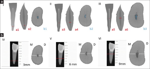
Materials and methods: Canal diameter and DT of mandibular incisors (n = 520) were evaluated using digital radiographs in buccolingual (BL) and mesiodistal (MD) directions. The diameter ratio (DR) BL/MD was classified: flattened (FL, DR >4); oval (OV, 2≤ DR ≥4); rounded (RN, 1.1< DR >2); round (RO, 0.9≤ DR ≥1.1); and with BL flatness (BL, DR <0.9). OV (n = 110) were subjected to micro-CT. DT and DR were evaluated at 3, 6, and 9 mm. ANOVA, Tukey, and paired Wilcoxon tests (P < 0.05) were used.
Results: Radiographic classification was 23.3% FL, 41.3% OV, 27.3% RN, 4.5% RO, and 3.6% BL. DT was similar. Radiographic DT at 3 and 9 mm was greater than micro-CT (P < 0.05) and was similar at 6 mm (P > 0.05). DR differed between the analyses. Oval canals were predominant at all levels radiographically and at 9 and 6 mm in micro-CT analysis, with greater variation at 3 mm.
Conclusion: Oval root canals are predominant in mandibular incisors at 9 mm. Radiographic DT is larger than observed in micro-CT at 3 and 9 mm, and the classification differed in each root level. The classification at 9 mm is indicated.
Keywords: Endodontics; X-ray microtomography; radiography; root canal anatomy.
Conflict of interest statement
There are no conflicts of interest.
Figures

References
-
- Lin Z, Hu Q, Wang T, Ge J, Liu S, Zhu M, et al. Use of CBCT to investigate the root canal morphology of mandibular incisors. Surg Radiol Anat. 2014;36:877–82. - PubMed
-
- Milanezi de Almeida M, Bernardineli N, Ordinola-Zapata R, Villas-Bôas MH, Amoroso-Silva PA, Brandão CG, et al. Micro-computed tomography analysis of the root canal anatomy and prevalence of oval canals in mandibular incisors. J Endod. 2013;39:1529–33. - PubMed
-
- Leoni GB, Versiani MA, Pécora JD, Damião de Sousa-Neto M. Micro-computed tomographic analysis of the root canal morphology of mandibular incisors. J Endod. 2014;40:710–6. - PubMed
-
- Freire LG, Gavini G, Cunha RS, Santos Md. Assessing apical transportation in curved canals: Comparison between cross-sections and micro-computed tomography. Braz Oral Res. 2012;26:222–7. - PubMed
-
- Vertucci FJ. Root canal anatomy of the mandibular anterior teeth. J Am Dent Assoc. 1974;89:369–71. - PubMed
LinkOut - more resources
Full Text Sources
Research Materials