. 2017 Nov 17;9(3):770-776.
doi: 10.1039/c7sc04014h. eCollection 2018 Jan 21.

Target-activated streptavidin-biotin controlled binding probe

Affiliations

Target-activated streptavidin-biotin controlled binding probe

Yung-Peng Wu et al. Chem Sci. .

Abstract

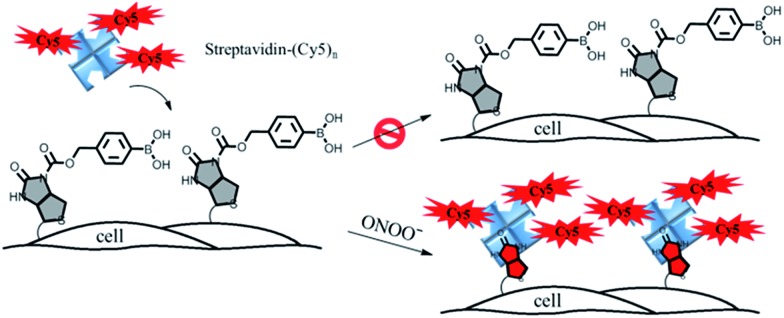
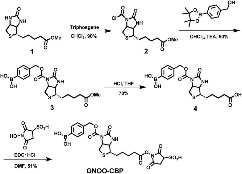
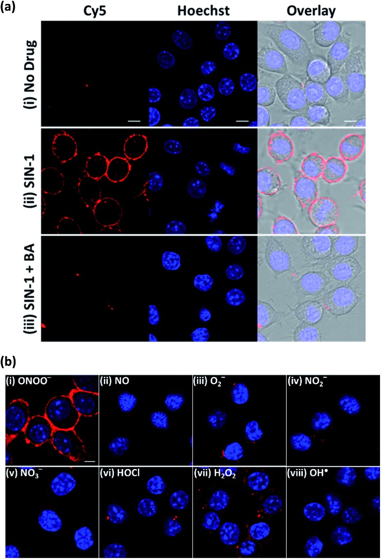
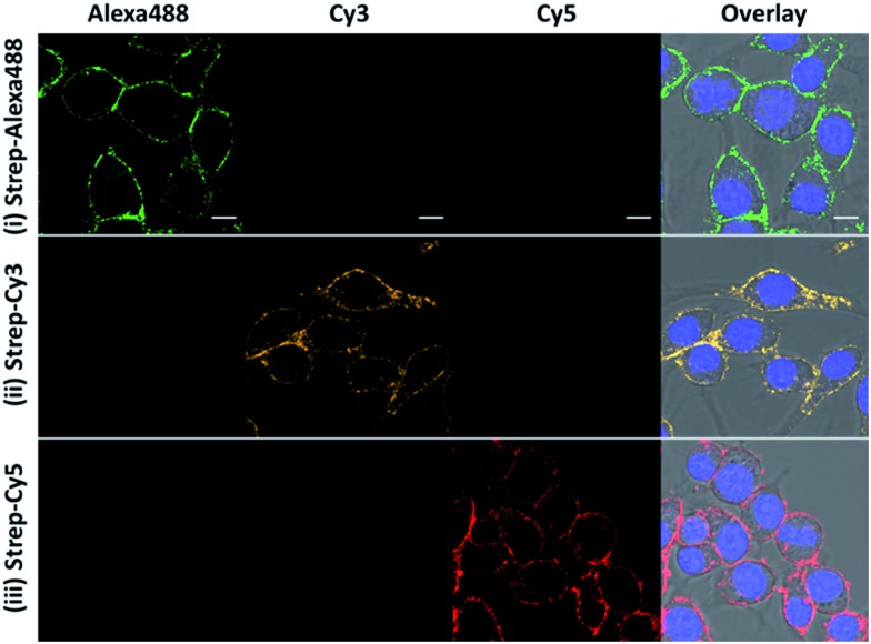
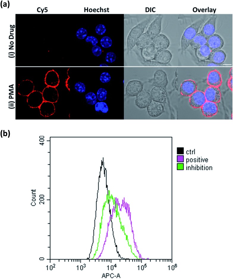
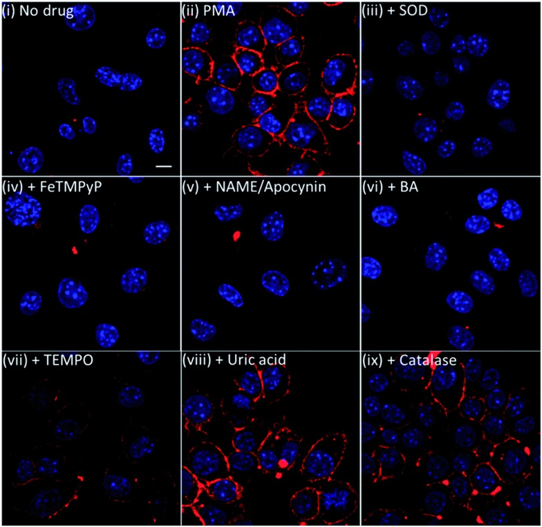
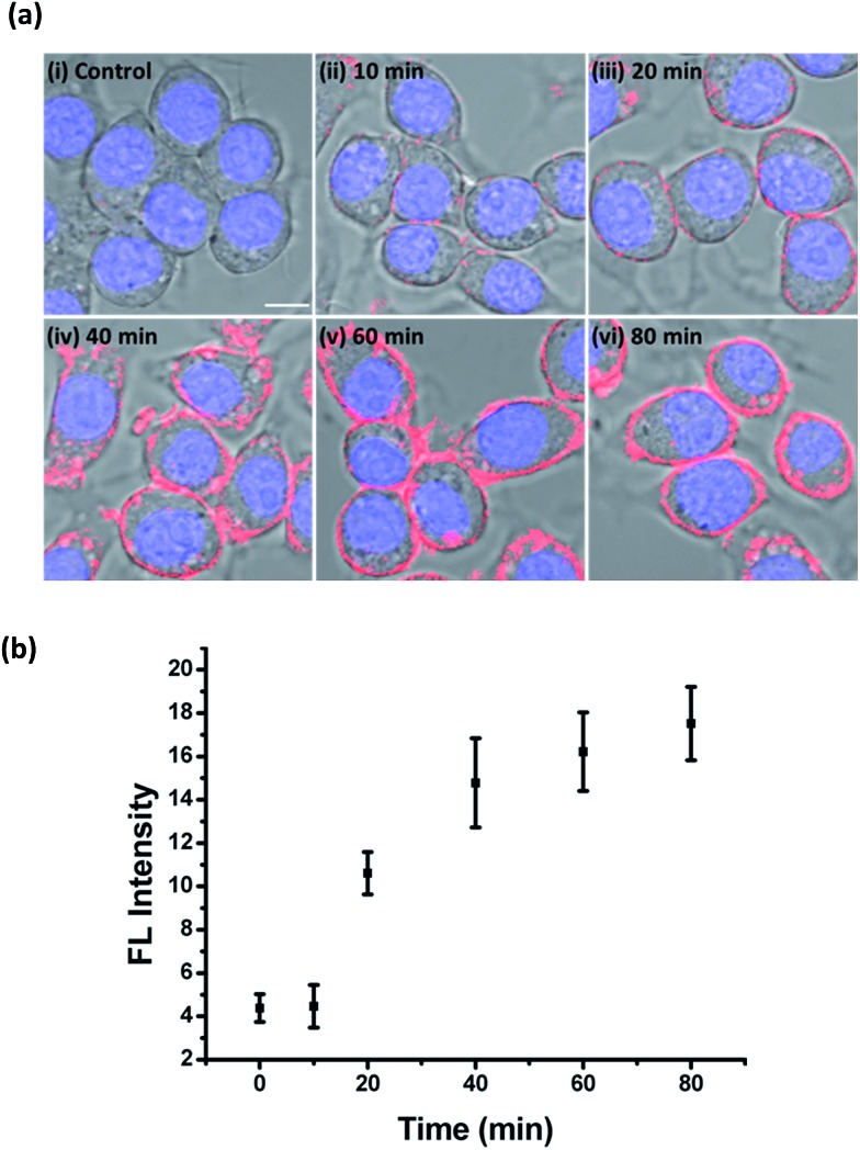
Target-activated chemical probes are important tools in basic biological research and medical diagnosis for monitoring enzyme activities and reactive small molecules. Based on the fluorescence turn-on mechanism, they can be divided into two classes: dye-based fluorescent probes and caged-luciferin. In this paper, we introduce a new type of chemical probe in which the fluorescence turn-on is based on controlled streptavidin-biotin binding. Compared to conventional probes, the streptavidin-biotin controlled binding probe has several advantages, such as minimal background at its "OFF" state, multiple signal amplification steps, and unlimited selection of the optimal dyes for detection. To expand the scope, a new synthetic method was developed, through which a wider range of analyte recognition groups can be easily introduced to construct the binding probe. This probe design was successfully applied to image and study secreted peroxynitrite (ONOO-) at the cell surface of macrophages where information on ONOO- is difficult to obtain. As the signals are generated upon the binding of streptavidin to the biotin probe, this highly versatile design can not only be used in fluorescence detection but can also be applied in various other detection modes, such as electrochemical and enzyme-amplified luminescence detection.


Figures

Fig. 1
Fig. 1. A schematic illustration of the membrane-anchored streptavidin–biotin controlled binding probe for the imaging of ONOO at the cell surface.
Scheme 1
Scheme 1. The synthesis of the cell-impermeable membrane-anchored streptavidin–biotin controlled binding probe, ONOO-CBP, for the detection of secreted ONOO at the extracellular surface.
Fig. 2
Fig. 2. The fluorescence response of the ONOO-CBP labeled RAW264.7 cells after incubation with different exogenous ROS and RNS for 60 minutes. (a) The live cell imaging of the ONOO-CBP labeled RAW264.7 cells in (i) the absence or (ii) the presence of 100 μM SIN-1 or (iii) 250 μM BA with 100 μM SIN-1. (b) The live cell imaging of the ONOO-CBP labeled RAW264.7 cells in the presence of (i) 50 μM SIN-1 or 100 μM each of (ii) NO, (iii) O2, (iv) NO2, (v) NO3, (vi) HOCl, (vii) H2O2, or (viii) OH˙. The cell surface stained with streptavidin–Cy5 is shown in red and the nuclei labeled with Hoechst 34580 are shown in blue. Scale bar: 20 μm.
Fig. 3
Fig. 3. The fluorescence response of the ONOO-CBP labeled RAW264.7 cells after incubation with 100 μM SIN-1 for 60 minutes. After biotin uncaging, the cells were stained with (i) streptavidin–Alexa488, (ii) streptavidin–Cy3, and (iii) streptavidin–Cy5. Scale bar: 20 μm.
Fig. 4
Fig. 4. (a) The live cell imaging of the ONOO-CBP labeled RAW264.7 cells in the (i) absence or (ii) presence of 1 μg mL–1 PMA after 60 minutes of incubation. Scale bar: 20 μm. (b) The fluorescence-activated cell sorting (FACS) analysis of the ONOO-CBP labeled RAW264.7 cells in the absence (black line) or presence (pink line) of 1 μg mL–1 PMA after 60 minutes of incubation. The green line indicates the condition in which 15 μg mL–1 SOD was mixed with 1 μg mL–1 PMA.
Fig. 5
Fig. 5. The live cell imaging of the ONOO-CBP labeled RAW264.7 cells in (i) the absence or (ii) the presence of 1 μg mL–1 PMA and (iii) PMA + 15 μg mL–1 SOD, (iv) PMA + 1 mM FeTMPyP, (v) PMA + 1 mM l-NAME and 1 mM apocynine, (vi) PMA + 250 μM BA, (vii) PMA + 1 mM TEMPO, (viii) PMA + 5 mM uric acid, or (ix) PMA + 10 μg mL–1 catalase. Besides the l-NAME/apocynin mixture, which was incubated with the cells for 90 minutes prior to PMA stimulation, all the inhibitors were added to the cells together with PMA and incubated for 60 minutes. The cells were stained with streptavidin–Cy5. Scale bar: 20 μm.
Fig. 6
Fig. 6. The time course of ONOO secretion into the extracellular medium upon PMA stimulation. (a) The live cell imaging of the ONOO-CBP labeled RAW264.7 cells in (i) the absence or presence of 1 μg mL–1 PMA after incubation for (ii) 10, (iii) 20, (iv) 40, (v) 60, or (vi) 80 minutes. Scale bar: 20 μm. (b) The time course of the mean fluorescence intensity (processed using Image J) on the cell surface of the RAW264.7 cells treated with 1 μg mL–1 PMA (N = 30 for each indicated time). The cells were stained with streptavidin–Cy5. Scale bar: 20 μm.

References

    1. Weissleder R. Nat. Biotechnol. 2001;19:316–317. - PubMed
    1. Johnson I., The Molecular Probes Handbook: A Guide to Fluorescent Probes and Labeling Technologies, Life Technologies Corporation, 11th edn, 2010.
    1. Xing B., Khanamiryan A., Rao J. J. Am. Chem. Soc. 2005;127:4158–4159. - PubMed
    1. Sakabe M., Asanuma D., Kamiya M., Iwatate R. J., Hanaoka K., Terai T., Nagano T., Urano Y. J. Am. Chem. Soc. 2012;135:409–414. - PubMed
    1. Lippert A. R., New E. J., Chang C. J. J. Am. Chem. Soc. 2011;133:10078–10080. - PubMed