

Validation of the UNC OCT Index for the Diagnosis of Early Glaucoma
- PMID: 29629238
- PMCID: PMC5886105
- DOI: 10.1167/tvst.7.2.16
Validation of the UNC OCT Index for the Diagnosis of Early Glaucoma
Abstract
Purpose: To independently validate the performance of the University of North Carolina Optical Coherence Tomography (UNC OCT) Index in diagnosing and predicting early glaucoma.
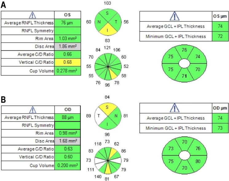
Methods: Data of 118 normal subjects (118 eyes) and 96 subjects (96 eyes) with early glaucoma defined as visual field mean deviation (MD) greater than -4 decibels (dB), aged 40 to 80 years, and who were enrolled in the Full-Threshold Testing Size III, V, VI comparison study were used in this study. CIRRUS OCT average and quadrants' retinal nerve fiber layer (RNFL); optic disc vertical cup-to-disc ratio (VCDR), cup-to-disc area ratio, and rim area; and average, minimum, and six sectoral ganglion cell-inner plexiform layer (GCIPL) measurements were run through the UNC OCT Index algorithm. Area under the receiver operating characteristic curve (AUC) and sensitivities at 95% and 99% specificity were calculated and compared between single parameters and the UNC OCT Index.
Results: Mean age was 60.1 ± 11.0 years for normal subjects and 66.5 ± 8.1 years for glaucoma patients (P < 0.001). MD was 0.29 ± 1.04 dB and -1.30 ± 1.35 dB in normal and glaucomatous eyes (P < 0.001), respectively. The AUC of the UNC OCT Index was 0.96. The best single metrics when compared to the UNC OCT Index were VCDR (0.93, P = 0.054), average RNFL (0.92, P = 0.014), and minimum GCIPL (0.91, P = 0.009). The sensitivities at 95% and 99% specificity were 85.4% and 76.0% (UNC OCT Index), 71.9% and 62.5% (VCDR, all P < 0.001), 64.6% and 53.1% (average RNFL, all P < 0.001), and 66.7% and 58.3% (minimum GCIPL, all P < 0.001), respectively.
Conclusions: The findings confirm that the UNC OCT Index may provide improved diagnostic perforce over that of single OCT parameters and may be a good tool for detection of early glaucoma.
Translational relevance: The UNC OCT Index algorithm may be incorporated easily into routine clinical practice and be useful for detecting early glaucoma.
Keywords: UNC OCT Index; early glaucoma; optical coherence tomography.
Figures


References
-
- Mwanza JC, Budenz DL, Godfrey DG,et al. . Diagnostic performance of optical coherence tomography ganglion cell–inner plexiform layer thickness measurements in early glaucoma. Ophthalmology. 2014; 121: 849– 854. - PubMed
-
- Mwanza JC, Durbin MK, Budenz DL,et al. . Glaucoma diagnostic accuracy of ganglion cell-inner plexiform layer thickness: comparison with nerve fiber layer and optic nerve head. Ophthalmology. 2012; 119: 1151– 1158. - PubMed
-
- Sung MS, Yoon JH, Park SW. . Diagnostic validity of macular ganglion cell-inner plexiform layer thickness deviation map algorithm using cirrus HD-OCT in preperimetric and early glaucoma. J Glaucoma. 2014; 23: e144– 151. - PubMed
Grants and funding
LinkOut - more resources
Full Text Sources
Other Literature Sources
Miscellaneous
