Case Reports
. 2018 Apr 9;33(15):e115.
doi: 10.3346/jkms.2018.33.e115.

Atypical Presentation of Pneumocystis jirovecii Infection in HIV Infected Patients: Three Different Manifestations

Affiliations
Case Reports

Atypical Presentation of Pneumocystis jirovecii Infection in HIV Infected Patients: Three Different Manifestations

Bongyoung Kim et al. J Korean Med Sci. .

Abstract

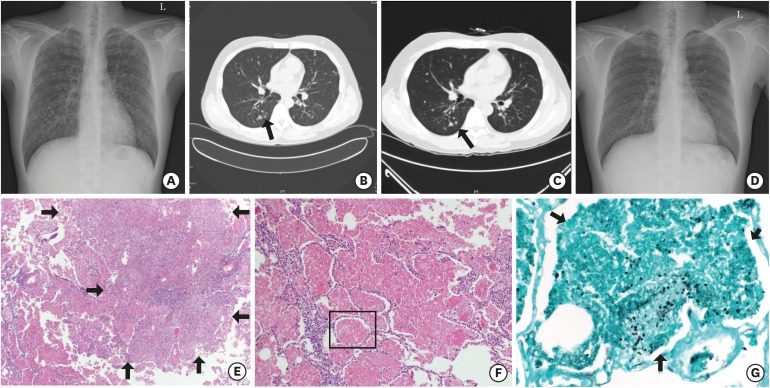
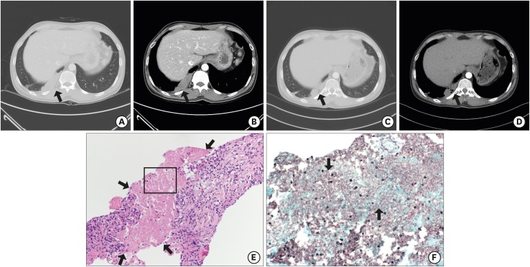
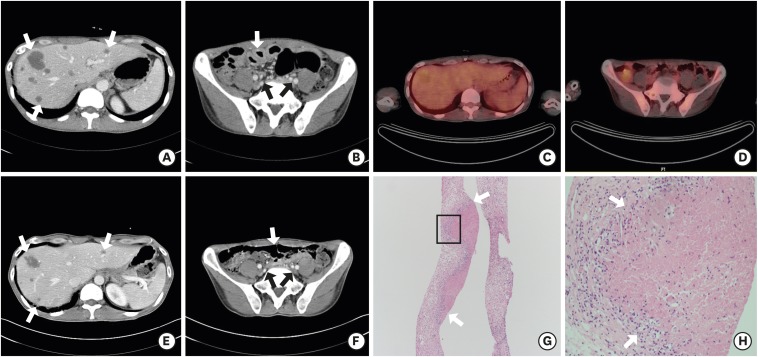
Advances in the treatment and prevention of Pneumocystis jirovecii infection (PJI) in human immunodeficiency virus (HIV) patients decreased incidence and mortality dramatically, however, it may be associated with an increased frequency of unusual manifestation such as cystic formation, pneumothorax, focal infiltration, nodular formation, or extrapulmonary lesions. We report three cases of PJI with atypical manifestations. Each case demonstrates different clinical features: multiple nodular pulmonary lesion (32-year-old man with abnormal chest X-ray finding), subpleural mass-like lesion (43-year-old man with left visual loss and right pleuritic chest pain), and extrapulmonary mass-like lesions in the liver, lymph nodes, and small bowel (39-year-old man with cough, sputum, and dyspnea). P. jirovecii was confirmed in all 3 cases and they were treated well. It is necessary to understand that PJI shows variable clinical features.

Keywords: AIDS; Atypical; Extrapulmonary; HIV; Nodular; Pneumocystis jirovecii.


Conflict of interest statement

The authors have no potential conflicts of interest to disclose.

Figures

Fig. 1
Fig. 1
Findings of the 32-year-old man with multiple pulmonary nodules (case 1). (A, B) Simple chest X-ray and chest CT on admission day show disseminated multiple nodules with some cavitation (arrow) on whole lung field. (C) Chest CT on hospital day 29 shows decreased size of multiple nodules with disappearance of cavitary lesions (arrow). (D) Simple chest X-ray after 16 months from discharge shows previous multiple nodules disappeared. (E-G) Histopathologic feature of lung specimen shows necrotizing granulomatous lesions (arrows) and pink, foamy exudate (E, F: H & E stain; × 100, × 200, respectively) containing oval shaped fungal cysts (arrows) within alveolar space (G: GMS stain; × 400). CT = computed tomography, H & E = hematoxylin and eosin, GMS = gomori methenamine-silver.
Fig. 2
Fig. 2
Findings of the 43-year-old man with subpleural mass-like lesion (case 2). (A, B) Chest CT on admission day shows a 13.35 cm2 lobulated homogeneously enhancing mass-like lesion (arrows) on right lower lobe. (C, D) Chest CT shows the mass size decreased to 5.87 cm2 (arrows) after one month from discharge. (E, F) Histopathologic feature of lung specimen shows fibrotic granulomatous lesions with necrotic amorphous bubbly area (arrows) (E: H & E stain; × 200) which contained oval shaped fungal cysts (arrows) (F: GMS stain; × 400). CT = computed tomography, H & E = hematoxylin and eosin, GMS = gomori methenamine-silver.
Fig. 3
Fig. 3
Findings of the 39-year-old man with extrapulmonary mass-like lesions (case 3). (A, B) Abdominal CT on admission day shows multiple hepatic nodules (arrows), multiple small lymph nodes in upper abdominal region (arrows), and diffuse jejunal wall thickening (arrows). (C, D) PET-CT on hospital day 10 shows no increased metabolic uptake on mentioned lesions, and diffuse jejunal wall thickening. (E, F) Abdominal CT on hospital day 17 shows significant improvement in mentioned areas (arrows). (G, H) Histopathologic feature of liver specimen shows granuloma with ill-formed necrosis (arrows) (H & E stain; × 200, × 400, respectively). CT = computed tomography, PET-CT = positron emission tomography-computed tomography, H & E = hematoxylin and eosin.

References

    1. The Korean Society for AIDS. Clinical guidelines for the treatment and prevention of opportunistic infections in HIV-infected Koreans. Infect Chemother. 2012;44(3):93–139. - PMC - PubMed
    1. Furrer H, Egger M, Opravil M, Bernasconi E, Hirschel B, Battegay M, et al. Discontinuation of primary prophylaxis against Pneumocystis carinii pneumonia in HIV-1 infected adults treated with combination antiretroviral therapy. Swiss HIV Cohort Study. N Engl J Med. 1999;340(17):1301–1306. - PubMed
    1. Kim JM, Cho GJ, Hong SK, Chang KH, Chung JS, Choi YH, et al. Epidemiology and clinical features of HIV infection/AIDS in Korea. Yonsei Med J. 2003;44(3):363–370. - PubMed
    1. Boiselle PM, Crans CA, Jr, Kaplan MA. The changing face of Pneumocystis carinii pneumonia in AIDS patients. AJR Am J Roentgenol. 1999;172(5):1301–1309. - PubMed
    1. Kim HS, Shin KE, Lee JH. Single nodular opacity of granulomatous Pneumocystis jirovecii pneumonia in an asymptomatic lymphoma patient. Korean J Radiol. 2015;16(2):440–443. - PMC - PubMed

Publication types