. 2019 Aug;39(8):1501-1515.
doi: 10.1177/0271678X18769515. Epub 2018 Apr 9.

PDGFR-β restores blood-brain barrier functions in a mouse model of focal cerebral ischemia

Affiliations

PDGFR-β restores blood-brain barrier functions in a mouse model of focal cerebral ischemia

Jie Shen et al. J Cereb Blood Flow Metab. 2019 Aug.

Abstract

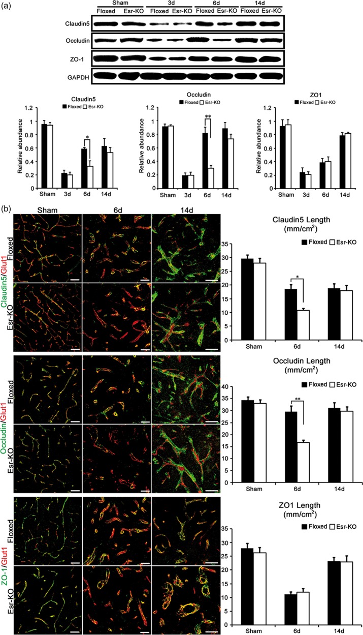
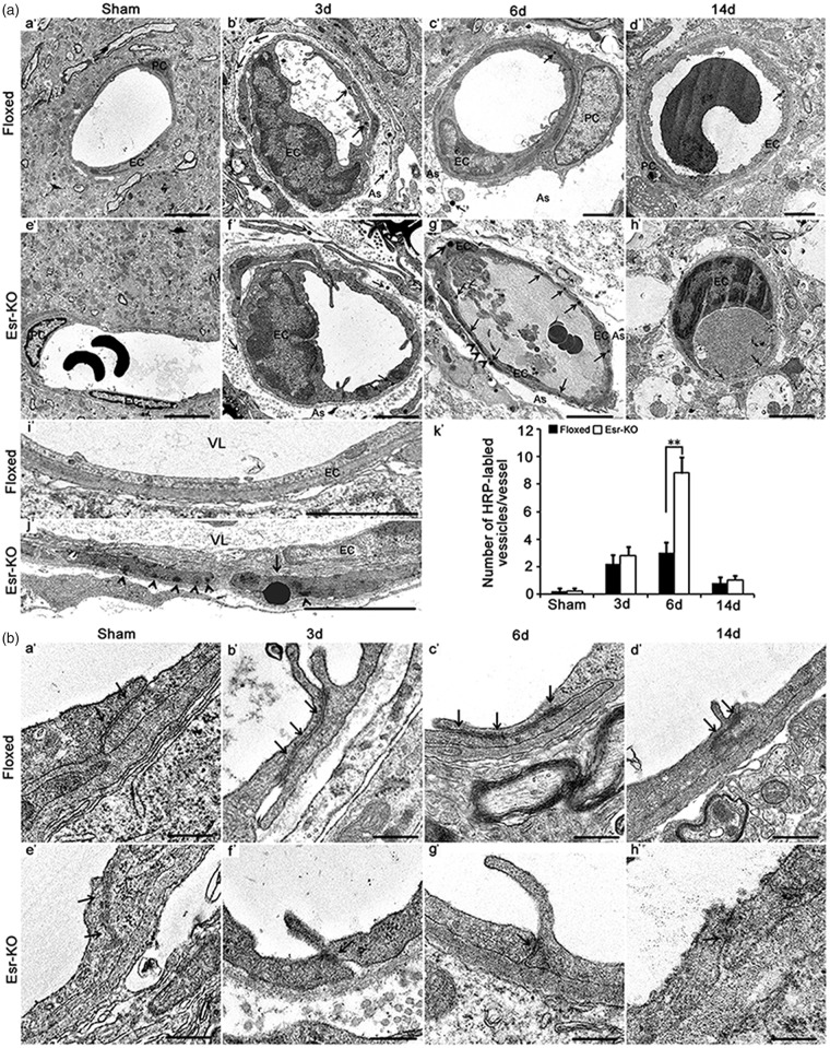
Although platelet-derived growth factor receptor beta (PDGFR-β) mediates the recruitment of vascular pericytes into ischemic lesion to restore the blood-brain barrier (BBB) dysfunction, its mechanisms still remain elusive. Compared with control PDGFR-βfloxed/floxed mice (Floxed), postnatally induced systemic PDGFR-β knockout mice (Esr-KO) not only showed severe brain edema, neurologic functional deficits, decreased expression of tight junction (TJ) proteins, abundant endothelial transcytosis, and deformed TJs in the BBB, but also showed reduced expression of transforming growth factor-β (TGF-β) protein after photothrombotic middle cerebral artery occlusion (MCAO). In endothelial-pericyte co-culture, an in vitro model of BBB, the increment in the barrier function of endothelial monolayer induced by pericyte co-culture was completely cancelled by silencing PDGFR-β gene expression in pericytes, and was additively improved by PDGFR-β and TGF-β receptor signals under hypoxia condition. Exogenous PDGF-BB increased the expression of p-Smad2/3, while anti-TGF-β1 antibody at least partially inhibited the phosphorylation of Smad2/3 after PDGF-BB treatment in vitro. Furthermore, pre-administration of TGF-β1 partially alleviated edema formation, neurologic dysfunction, and TJs reduction in Esr-KO mice after MCAO. Accordingly, PDGFR-β signalling, via TGF-β signalling, may be crucial for restoration of BBB integrity after cerebral ischemia and therefore represents a novel potential therapeutic target.

Keywords: Blood-brain barrier; cerebral ischemia; pericyte; platelet-derived growth factor receptor-beta; transforming growth factor-β.


Figures

Figure 1.
Figure 1.
PDGFR-β deletion exacerbated edema formation and impaired functional recovery after cerebral ischemia. (a) Absolute brain water content, edema formation and space-occupying effect of edema in control mice (Floxed, n = 7) and PDGFR-β knockout mice (Esr-KO, n = 7) from 3 to 14 days after MCAO and sham-operated mice. (b) Adhesive removal test performances. Contralesional contact time and removal time, ipsilesional contact time and removal time in Floxed (n = 7) and Esr-KO (n = 7) mice before and after MCAO. (c) Percentage of right turns in Floxed (n = 7) and Esr-KO (n = 7) mice before and after MCAO. *P < 0.05, **P < 0.01.
Figure 2.
Figure 2.
PDGFR-β deletion suppressed the recovery of tight junction proteins after cerebral ischemia. (a) Western blot analysis and quantification of Claudin5, Occludin, and ZO-1 protein expression in ipsilesional cerebral cortices of Floxed and Esr-KO mice after MCAO and sham-operated mice (n = 3 per group). Each bar was normalised by GAPDH. (b) Double-immunofluorescence staining for Claudin5 (green)/Glut1 (red), Occludin (green)/Glut1 (red) and ZO-1 (green)/Glut1 (red), and quantification of Claudin5, Occludin and ZO-1 length per area in the ischemic border of cerebral cortices of Floxed and Esr-KO mice at 6 and 14 days after MCAO and cerebral cortices of sham-operated mice (sham), n = 6 per group. Scale bars = 50 µm. *P < 0.05, **P < 0.01.
Figure 3.
Figure 3.
PDGFR-β deletion increased transcellular transport of deformation of tight junctions after cerebral ischemia. (a) Electron microscopy images and quantification of intravenously injected tracer-HRP labelled vesicles in the vascular endothelium in the ischemic border of Floxed and Esr-KO mice from 3 to 14 days after MCAO (b′–d′, f′–h′), and sham-operated mice (sham; n = 6 per group, a′, e′). (i′, j′): Electron microscopy images at higher magnification of intravenously injected tracer-HRP labelled vesicles in the vascular endothelium in the ischemic border of Floxed and Esr-KO mice at six days after MCAO. PC: pericyte, EC: endothelial cell; As: astrocyte; VL: vascular lumen; arrows indicate HRP-labelled vascular vesicles; arrowheads indicate HRP accumulation in basal membrane. Scale bars = 2 µm in Figure a′ to h′, 10 µm in (i′ and j′). (k′) Quantification of number of HRP-labelled vascular vesicles per vessel in the ischemic border of Floxed and Esr-KO mice from 3 to 14 days after MCAO and sham-operated mice (sham; n = 6 per group). *P < 0.05, **P < 0.01. (b) Deformation of tight junctions in the vascular endothelium in the ischemic border of Floxed and Esr-KO mice from 3 to 14 days after MCAO, and sham-operated mice (sham), n = 6 per group. Arrows indicate tight junctions. Scale bars = 400 nm.
Figure 4.
Figure 4.
PDGFR-β deletion reduced expression of TGF-β protein after cerebral ischemia. (a) Western blot analysis and quantification of TGF-β, Angiopiontin 1(Ang 1), Tie2, Aquaporin 4 (Aqp 4) protein expressions in ipsilesional cerebral cortices of Floxed and Esr-KO mice after MCAO and sham-operated mice (n = 3 per group). Each bar was normalised by GAPDH. *P < 0.05, **P < 0.01. (b) Quadruple immunofluorescence staining for TGF-β (green)/Desmin (purple)/Glut1 (red) in the ischemic border of cerebral cortices of Floxed and Esr-KO mice at six days after MCAO. Counterstained with DAPI (blue). n = 6 per group. Scale bars = 10 µm. (c) Triple immunofluorescence staining for Aqp 4 (green)/Glut1 (red) and GFAP (green)/Glut1 (red) in the ischemic border of cerebral cortices of Floxed and Esr-KO mice at 6 and 14 days after MCAO and cerebral cortices of sham-operated mice (sham). Counterstained with DAPI (blue). n = 6 per group. Scale bars = 10 µm.
Figure 5.
Figure 5.
PDGFR-β signalling regulated BBB permeability via TGF-β in vitro and in vivo. (a) In in vitro co-culture model, quantitative real-time PCR of PDGFR-β mRNA with GAPDH siRNA, Negative control (NC) siRNA, three PDGFR-β siRNAs transfection or non-transfection (vehicle). (b) MTT assay and (c) TUNEL staining and percentage TUNEL-positive pericytes from in vitro co-culture model with three PDGFR-β siRNAs transfection or non-transfection (vehicle). (d) Exogenous TGF-β (1 ng/ml) treatment for permeability coefficients of 14C-sucrose of HBMVECs alone (EC) and co-culture model (Co) and co-culture model with PDGFR-β siRNA knockdown (Co-KO), or no-treatment. SB431542 treatment (a selective TGF-β type I receptor inhibitor) for permeability coefficients of 14C-sucrose of HBMVECs alone (EC) and co-culture model (Co) and co-culture model with PDGFR-β siRNA knockdown (Co-KO), or no-treatment. (e) Western blot analysis and quantification of p-Smad2/3 protein expression with PDGF-BB treatment and/or anti-TGF-β1 antibody treatment in co-culture model. (f) Edema formation, (g) contralesional contact time and removal time in control mice (Floxed, n = 7) and PDGFR-β knockout mice (Esr-KO, n = 7) at six days after MCAO with or without pre-administration of TGF-β1. (h) Western blot analysis and quantification of Claudin5, Occludin, and ZO1 protein expression in ipsilesional cerebral cortices of Floxed and Esr-KO mice (n = 3 per group) at 6 days after MCAO with or without pre-administration of TGF-β1. *P < 0.05, **P < 0.01.

References

    1. Prakash R, Carmichael ST. Blood-brain barrier breakdown and neovascularization processes after stroke and traumatic brain injury. Curr Opin Neurol 2015; 28: 556–564. - PMC - PubMed
    1. Zhao Z, Nelson AR, Betsholtz C, et al. Establishment and dysfunction of the blood-brain barrier. Cell 2015; 163: 1064–1078. - PMC - PubMed
    1. Cai W, Liu H, Zhao J, et al. Pericytes in brain injury and repair after ischemic stroke. Transl Stroke Res 2017; 8: 107–121. - PMC - PubMed
    1. Tallquist M, Kazlauskas A. PDGF signalling in cells and mice. Cytokine Growth Factor Rev 2004; 15: 205–213. - PubMed
    1. Sweeney MD, Ayyadurai S, Zlokovic BV. Pericytes of the neurovascular unit: key functions and signalling pathways. Nat Neurosci 2016; 19: 771–783. - PMC - PubMed

Publication types

MeSH terms

Substances

LinkOut - more resources