

doi: 10.1038/nmeth.4661.
Epub 2018 Apr 9.
Real-time 3D single-molecule localization using experimental point spread functions
Affiliations
- PMID: 29630062
- PMCID: PMC6009849
- DOI: 10.1038/nmeth.4661
Item in Clipboard
Real-time 3D single-molecule localization using experimental point spread functions
Nat Methods.
2018 May.
Abstract
We present a real-time fitter for 3D single-molecule localization microscopy using experimental point spread functions (PSFs) that achieves minimal uncertainty in 3D on any microscope and is compatible with any PSF engineering approach. We used this method to image cellular structures and attained unprecedented image quality for astigmatic PSFs. The fitter compensates for most optical aberrations and makes accurate 3D super-resolution microscopy broadly accessible, even on standard microscopes without dedicated 3D optics.
Conflict of interest statement
The authors declare no competing financial interests.
Figures
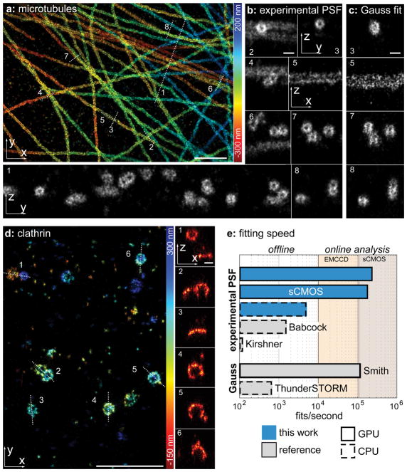
(a) Immunolabeled microtubules imaged using the DNA-PAINT approach. Localizations are color-coded according to their z-position. Corresponding localization precisions and profiles are shown in Supplementary Fig. 4. (b) Side-view cross-sections along the lines denoted in (a) clearly reveal the hollow, cylinder-shaped structure of microtubules. (c) Side-view reconstructions of the same area as in (b) analysed with ThunderSTORM using an elliptical Gaussian MLE fit. (d) Immunolabeled clathrin imaged using dSTORM. Side-view cross-sections clearly show the geometry of clathrin-coated pits with low and high curvatures. (e) Fitting speed of the fitter presented in this work, compared to previous implementation of fitters for experimental PSF models (Babcock et al. and Kirshner et al.) and Gaussian PSF models (Smith et al. and ThunderSTORM). Fits/second were measured on a i7-5930 CPU and a GTX1070 consumer graphics card. Width of the cross-sections: 150 nm (b1,4,6), 200 nm (b2,3,7,8), 30 nm (b5), 50 nm (d). Scale bars: 1 μm (a, d) and 100 nm (b, c, and x-z reconstructions in d).
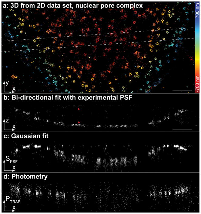
(a) Nup107-SNAP-AlexaFluor647 was imaged using dSTORM on a standard microscope without 3D optics. (b) Side-view reconstruction of the region denoted in (a). The nucleoplasmic and cytoplasmic rings of the nuclear pore complex, spaced 53 nm apart, can be easily resolved. The arrows denote nuclear pore complexes and their mirror images caused by misassignments. From the respective number of localizations, we estimated the fraction of misassignments to be ~5%. (c) Side view reconstruction of the same region using the size of the PSF SPSF from a fit with a symmetric Gaussian PSF model as a measure for the z-position. (d) Side view reconstruction of the same region using the photometry-based intensity ratio PTRABI as a measure for the z-position. Corresponding localization precisions and profiles can be found in Supplementary Fig. 4. Scale bars: 1 μm.
References
-
- Baddeley D, Cannell MB, Soeller C. Three-dimensional sub-100 nm super-resolution imaging of biological samples using a phase ramp in the objective pupil. Nano Res. 2011;4:589–598.
-
- Juette MF, et al. Three-dimensional sub–100 nm resolution fluorescence microscopy of thick samples. Nat Methods. 2008;5:527–529. - PubMed
Publication types
MeSH terms
Grants and funding
LinkOut - more resources
Full Text Sources
Other Literature Sources
Research Materials
