Review
. 2018 Sep;91(1089):20170762.
doi: 10.1259/bjr.20170762. Epub 2018 Apr 20.

The importance of the vascular endothelial barrier in the immune-inflammatory response induced by radiotherapy

Affiliations
Review

The importance of the vascular endothelial barrier in the immune-inflammatory response induced by radiotherapy

Olivier Guipaud et al. Br J Radiol. 2018 Sep.

Abstract

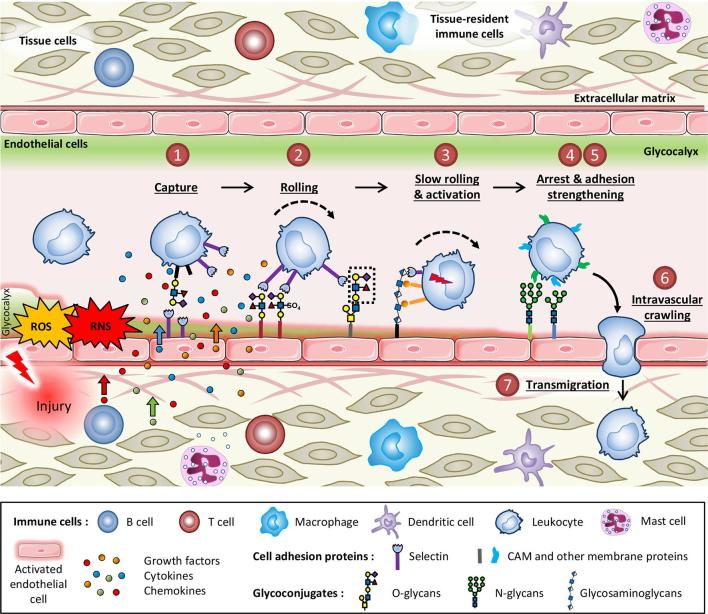
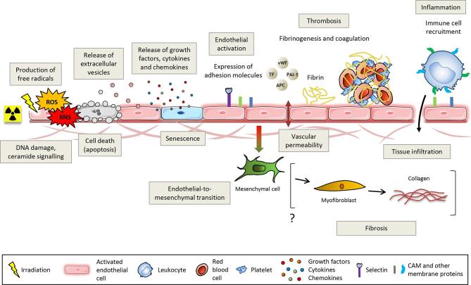
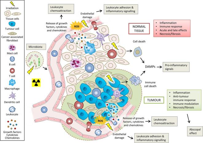
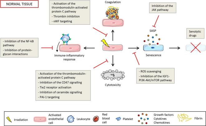
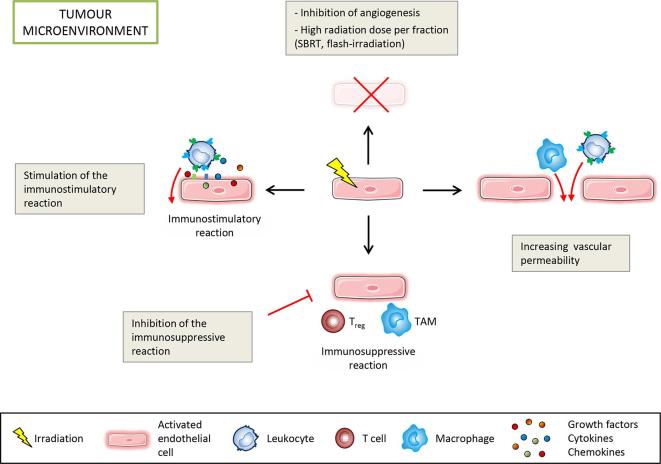
Altered by ionising radiation, the vascular network is considered as a prime target to limit normal tissue damage and improve tumour control in radiotherapy (RT). Irradiation damages and/or activates endothelial cells, which then participate in the recruitment of circulating cells, especially by overexpressing cell adhesion molecules, but also by other as yet unknown mechanisms. Radiation-induced lesions are associated with infiltration of immune-inflammatory cells from the blood and/or the lymph circulation. Damaged cells from the tissues and immune-inflammatory resident cells release factors that attract cells from the circulation, leading to the restoration of tissue balance by fighting against infection, elimination of damaged cells and healing of the injured area. In normal tissues that surround the tumours, the development of an immune-inflammatory reaction in response to radiation-induced tissue injury can turn out to be chronic and deleterious for the organ concerned, potentially leading to fibrosis and/or necrosis of the irradiated area. Similarly, tumours can elicit an immune-inflammation reaction, which can be initialised and amplified by cancer therapy such as radiotherapy, although immune checkpoints often allow many cancers to be protected by inhibiting the T-cell signal. Herein, we have explored the involvement of vascular endothelium in the fate of healthy tissues and tumours undergoing radiotherapy. This review also covers current investigations that take advantage of the radiation-induced response of the vasculature to spare healthy tissue and/or target tumours better.


Figures

Figure 1.
Figure 1.
The leukocyte adhesion cascade is triggered by a pro-inflammatory stimulus. Injury activates endothelial cells, allows production of free radicals and damages tissue and tissue-resident immune cells leading to the release of cytokines, chemokines and growth factors, which then attract leukocytes from the circulation. Circulating leukocytes undertake a seven-step process of capture, rolling, slow rolling and activation, arrest and adhesion strengthening, intravascular crawling and finally transmigration to reach sites of inflammation. Each step of this process is controlled by various adhesion molecules at the surface of the endothelium. All of these proteins are glycosylated, a post-translational modification process that may be regulated during inflammation. During this cascade of events, leukocytes are also activated by interactions with cytokines, chemokines and growth factors sequestrated by the glycosaminoglycans of the endothelial glycocalyx. RNS, reactive nitrogen species; ROS, reactive oxygen species.
Figure 2.
Figure 2.
Ionising radiation injures the vascular endothelium. DNA damage and ceramide production lead to cell death, stress-induced premature senescence, cell activation mainly characterised by the overexpression of adhesion molecules and disruption of the endothelial barrier. Endothelial activation promotes a pro-thrombotic and pro-inflammatory phenotype that ultimately leads to thrombosis and recruitment of leukocytes. Irradiated endothelial cells can also undergo an endothelial-to-mesenchymal transition that potentially contributes to fibrosis via a final differentiation into activated myofibroblasts capable of secreting collagens. APC, antigen-presenting cells; CAM, cell adhesion molecules; PAI-1, plasminogen activator inhibitor-type 1; TF, tissue factor; vWF, von Willebrand factor.
Figure 3.
Figure 3.
Immune-inflammatory effects of radiation exposure on normal tissues, tumours and tumour microenvironment. Ionising radiation causes tumour and normal cell damage, cell death and release of DAMPS, which act as pro-inflammatory signals. Radiation activates cells that then release cytokines/chemokines. Activated endothelial cells acquire a pro-inflammatory phenotype which promotes the leukocyte adhesion cascade. These initial responses finally lead to the recruitment and activation of diverse immune cells, which can then also participate in the abscopal effect of radiotherapy. Delayed mitotic death and proliferation of immune cells in the tissue then cause environmental changes that ultimately contribute to necrosis or fibrosis. Irradiation of the microbiota of the intestinal tract can also influence the responses of normal tissues and tumours through immune-vascular crosstalk. DAMPS, damage-associated molecular patterns; ROS, reactive oxygen species.
Figure 4.
Figure 4.
Potential endothelial-oriented strategies to spare normal tissue by targeting cytotoxicity, coagulation, activation of the immune-inflammatory response and senescence. PAI-1, plasminogen activator inhibitor-type 1; ROS, reactive oxygen species; SASP, senescence-associated secretory phenotype; vWF, von Willebrand factor.
Figure 5.
Figure 5.
Endothelial-oriented strategies to enhance tumour control by targeting the immunostimulatory and immunosuppressive reactions, inducing cell death and increasing vascular permeability to allow immune cell infiltration. SBRT, stereotactic body radiation therapy, TAM, tumour-associated macrophage.

References

    1. Shrieve DC, Loeffler JS. Human radiation injury. Philadelphia: The British Institute of Radiology.; 2011.
    1. Stone HB, McBride WH, Coleman CN. Modifying normal tissue damage postirradiation. Report of a workshop sponsored by the Radiation Research Program, National Cancer Institute, Bethesda, Maryland, September 6-8, 2000. Radiat Res 2002; 157: 204–23. - PubMed
    1. DeVita VT, Lawrence TS, Rosenberg SA. DeVita, Hellman, and Rosenberg’s cancer: principles & practice of oncology: The British Institute of Radiology.; 2011.
    1. Moding EJ, Kastan MB, Kirsch DG. Strategies for optimizing the response of cancer and normal tissues to radiation. Nat Rev Drug Discov 2013; 12: 526–42. doi: 10.1038/nrd4003 - DOI - PMC - PubMed
    1. Begg AC, Stewart FA, Vens C. Strategies to improve radiotherapy with targeted drugs. Nat Rev Cancer 2011; 11: 239–53. doi: 10.1038/nrc3007 - DOI - PubMed

MeSH terms