Comparative Study
. 2018 Oct;68(4):1347-1360.
doi: 10.1002/hep.29914. Epub 2018 Jul 16.

Neutrophil extracellular traps promote inflammation and development of hepatocellular carcinoma in nonalcoholic steatohepatitis

Affiliations
Comparative Study

Neutrophil extracellular traps promote inflammation and development of hepatocellular carcinoma in nonalcoholic steatohepatitis

Dirk J van der Windt et al. Hepatology. 2018 Oct.

Abstract

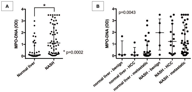
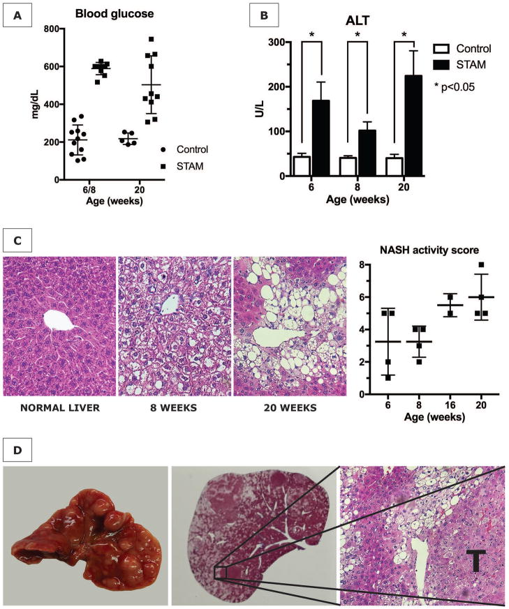
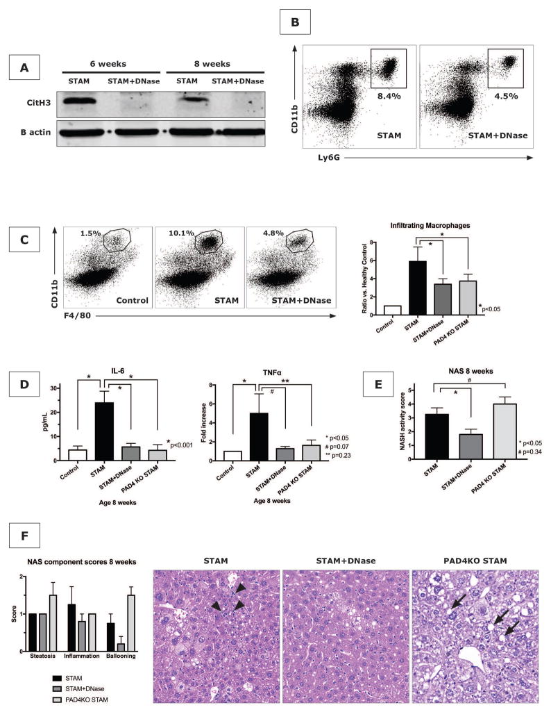
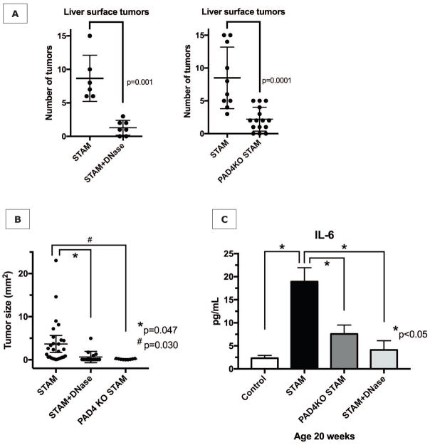
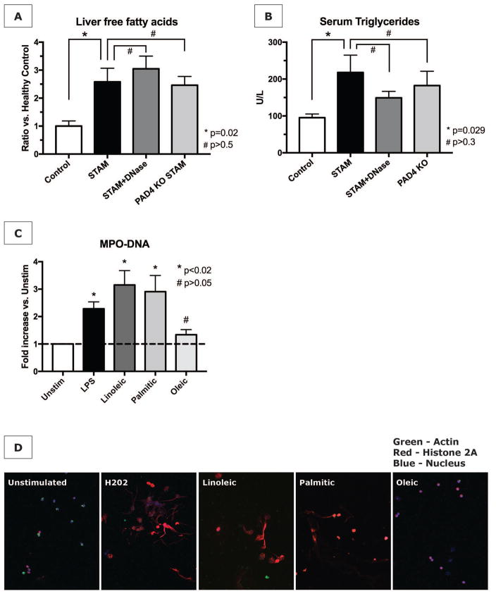
Nonalcoholic steatohepatitis (NASH) is a progressive, inflammatory form of fatty liver disease. It is the most rapidly rising risk factor for the development of hepatocellular carcinoma (HCC), which can arise in NASH with or without cirrhosis. The inflammatory signals promoting the progression of NASH to HCC remain largely unknown. The propensity of neutrophils to expel decondensed chromatin embedded with inflammatory proteins, known as neutrophil extracellular traps (NETs), has been shown to be important in chronic inflammatory conditions and in cancer progression. In this study, we asked whether NET formation occurs in NASH and contributes to the progression of HCC. We found elevated levels of a NET marker in serum of patients with NASH. In livers from STAM mice (NASH induced by neonatal streptozotocin and high-fat diet), early neutrophil infiltration and NET formation were seen, followed by an influx of monocyte-derived macrophages, production of inflammatory cytokines, and progression of HCC. Inhibiting NET formation, through treatment with deoxyribonuclease (DNase) or using mice knocked out for peptidyl arginine deaminase type IV (PAD4-/- ), did not affect the development of a fatty liver but altered the consequent pattern of liver inflammation, which ultimately resulted in decreased tumor growth. Mechanistically, we found that commonly elevated free fatty acids stimulate NET formation in vitro.

Conclusion: Our findings implicate NETs in the protumorigenic inflammatory environment in NASH, suggesting that their elimination may reduce the progression of liver cancer in NASH. (Hepatology 2018).


Conflict of interest statement

The authors declare that no conflict of interest exists

Figures

Figure 1
Figure 1. Increased NET marker in human NASH
(A) Patients with histopathologically proven NASH (n=46) have increased serum levels of MPO-DNA when compared with matched patients with normal liver histology (n=30). (B) MPO-DNA levels stratified by disease type (benign, primary malignant, metastatic). For any disease type, median MPO-DNA levels were higher when NASH was present in the background liver compared to a normal background liver.
Figure 2
Figure 2. The STAM model of diabetes and high fat diet leading to NASH and HCC
(A) STAM mice are consistently hyperglycemic, (B) have elevated serum alanine aminotransferase (ALT) levels, and (C) develop steatosis with progressively increasing NASH activity scores (NAS). (D) By 20 weeks, male mice develop numerous liver tumors (T) with characteristics of hepatocellular carcinoma in the fatty liver background.
Figure 3
Figure 3. The STAM model of NASH results in neutrophil infiltration and NET formation prior to attraction of macrophages
(A and B) STAM mice demonstrate an increase in infiltrating neutrophils between 5 and 12 weeks in the liver starting at 5 weeks. Because healthy control mice demonstrated a decline in hepatic neutrophils during the first 2 months of life, neutrophil count can be expressed as a ratio of STAM/control mice. (C) Western blot of livers from STAM mice have elevated levels of the NET marker citrullinated histone 3 (CitH3), compared to age-matched healthy controls (HC), which persists throughout the full course of the experiment. On quantification for age 8 weeks (n=4 in each group), CitH3 was significantly increased in STAM mice. (D) By 6 weeks of age, GFP-positive infiltrates (green) are detectable in STAM livers (middle panel, scale bar 100μm), compared to healthy control in left panel, with abundant presence of CitH3 immunofluorescent staining (red) and neutrophils with typical NET morphology (right panel, scale bar 40μm). Blue = nucleus. Grey = actin. (E) Infiltrating macrophages infiltrate the livers of STAM mice at a later time point (8 weeks) than neutrophils, concurrent with an increase in the serum level of IL-6 (F).
Figure 4
Figure 4. NET inhibition reduces macrophage infiltration, inflammatory cytokines, and NASH
(A) DNase treatment reduces liver CitH3 expression in STAM mice. (B) NET blockade with DNase in STAM mice reduces further neutrophil infiltration as shown for 8 weeks. Both DNase and PAD4−/− reduced several hallmarks of NASH including macrophage infiltration (n=4 mice per group) (C) and production of inflammatory cytokines IL-6 and TNF-α (n=8 mice per group) (D). NASH activity scores (n=5 mice per group) (E), with a reduction of inflammation, but unaffected presence of liver fat (F). H&E stained liver of STAM mice with lobular inflammation (arrow heads), which was improved by DNase treatment and in PAD4−/− mice, although PAD4−/− livers demonstrated accelerated development of steatosis (arrows) (F). (all shown for time point 8 weeks).
Figure 5
Figure 5. NET inhibition reduces progression of NASH to HCC
(A) NET inhibition for 20 weeks with daily DNase treatment, or by gene knockout for peptidylarginine deaminase (PAD4KO) significant reduces tumor formation at 20 weeks. (B) Tumor size analysis of the largest 3 tumors found at a random section through the liver revealed significantly reduced tumor sizes in DNase-treated mice and PAD4−/− mice, compared to untreated wild type STAM mice. (C) Serum IL-6 levels remain decreased, suggesting that NET inhibition persistently alters the inflammatory environment in which HCC arises.
Figure 6
Figure 6. Free fatty acids stimulate NET formation in vitro
(A) Livers from STAM mice have increased levels of free fatty acids (FFA). NET inhibition does not prevent the increase in FFA. (B) NET abrogation also does not prevent elevated serum triglycerides. (C) Isolated neutrophils were stimulated with LPS as positive control, or with various FFA at 50nM for 4h. MPO-DNA complexes in culture supernatants were significantly elevated after stimulation with linoleic and palmitic acid (data represent n=4 independent experiments per group). (D) Immunofluorescent staining for NETs was concordant with supernatant MPO-DNA levels.

References

    1. Loomba R, Sanyal AJ. The global NAFLD epidemic. Nat Rev Gastroenterol Hepatol. 2013;10:686–690. - PubMed
    1. Wong VW, Chitturi S, Wong GL, Yu J, Chan HL, Farrell GC. Pathogenesis and novel treatment options for non-alcoholic steatohepatitis. Lancet Gastroenterol Hepatol. 2016;1:56–67. - PubMed
    1. Mittal S, El-Serag HB, Sada YH, Kanwal F, Duan Z, Temple S, May SB, et al. Hepatocellular Carcinoma in the Absence of Cirrhosis in United States Veterans is Associated With Nonalcoholic Fatty Liver Disease. Clin Gastroenterol Hepatol. 2016;14:124–131. e121. - PMC - PubMed
    1. Hanahan D, Weinberg RA. Hallmarks of cancer: the next generation. Cell. 2011;144:646–674. - PubMed
    1. Gao B, Tsukamoto H. Inflammation in Alcoholic and Nonalcoholic Fatty Liver Disease: Friend or Foe? Gastroenterology. 2016;150:1704–1709. - PMC - PubMed

Publication types

MeSH terms