. 2018 Aug 1;315(2):F323-F331.
doi: 10.1152/ajprenal.00495.2017. Epub 2018 Apr 4.

Loss of biliverdin reductase-A promotes lipid accumulation and lipotoxicity in mouse proximal tubule cells

Affiliations

Loss of biliverdin reductase-A promotes lipid accumulation and lipotoxicity in mouse proximal tubule cells

Samuel O Adeosun et al. Am J Physiol Renal Physiol. .

Abstract

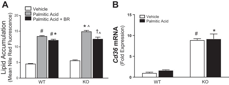
Obesity and increased lipid availability have been implicated in the development and progression of chronic kidney disease. One of the major sites of renal lipid accumulation is in the proximal tubule cells of the kidney, suggesting that these cells may be susceptible to lipotoxicity. We previously demonstrated that loss of hepatic biliverdin reductase A (BVRA) causes fat accumulation in livers of mice on a high-fat diet. To determine the role of BVRA in mouse proximal tubule cells, we generated a CRISPR targeting BVRA for a knockout in mouse proximal tubule cells (BVRA KO). The BVRA KO cells had significantly less metabolic potential and mitochondrial respiration, which was exacerbated by treatment with palmitic acid, a saturated fatty acid. The BVRA KO cells also showed increased intracellular triglycerides which were associated with higher fatty acid uptake gene cluster of differentiation 36 as well as increased de novo lipogenesis as measured by higher neutral lipids. Additionally, neutrophil gelatinase-associated lipocalin 1 expression, annexin-V FITC staining, and lactate dehydrogenase assays all demonstrated that BVRA KO cells are more sensitive to palmitic acid-induced lipotoxicity than wild-type cells. Phosphorylation of BAD which plays a role in cell survival pathways, was significantly reduced in palmitic acid-treated BVRA KO cells. These data demonstrate the protective role of BVRA in proximal tubule cells against saturated fatty acid-induced lipotoxicity and suggest that activating BVRA could provide a benefit in protecting from obesity-induced kidney injury.

Keywords: CRISPR; apoptosis; bilirubin; kidney; obesity; palmitic acid.


Figures

Fig. 1.
Fig. 1.
Characterization of biliverdin reductase A (BVRA)-deficient mouse proximal tubule cells. A. genotyping scheme of CRISPR-Cas9 double targeted BVRA knockout (KO) cells. PCR primers were designed outside of exon 1 and in exon 5 to detect a 316 bp fragment (normally 16,513 bp) in double mutant cells. Primers were also designed in exon 2 and exon 3 to generate a control, 706-bp PCR product. B. real time PCR expression of biliverdin reductase A gene (Blvra) mRNA in wild-type (WT) (open bar) and KO (closed bar) cells, n = 3 per group. C. Western blot of BVRA protein in WT and KO cells, n = 3 per group. D. bilirubin concentration in WT and KO cells under basal conditions and 21 h after treatment with biliverdin (40 µmol/l). *P < 0.05 as compared with WT. #P < 0.05 as compared with WT vehicle. ^P < 0.05 as compared with biliverdin treated WT. HSP90, heat shock protein 90.
Fig. 2.
Fig. 2.
The loss of biliverdin reductase A (BVRA) changes the mitochondrial respiration and metabolic potential of mouse proximal tubule cells. A. oxygen consumption rate (OCR) and extracellular acidification rate (ECAR) were used to quantify the cellular energy phenotype of wild-type (WT) and BVRA knockout (KO) mouse proximal tubule (MCT) cells with and without palmitic acid (400 μM) treatment. B. metabolic potential measured as the percentage increase of stressed OCR over baseline OCR and stressed ECAR over baseline ECAR were analyzed in the WT and BVRA KO MCT cells with and without palmitic acid (400 μM) treatment. C. mitochondrial respiration rates of the WT and BVRA KO cells were measured by OCR. D: the rate of glycolysis of the WT and BVRA KO cells was measured by the ECAR. *P < 0.05 as compared with WT vehicle. #P < 0.05 as compared with palmitic acid-treated WT. ^P < 0.05 as compared with KO vehicle. N = 10–20 per group.
Fig. 3.
Fig. 3.
Effect of biliverdin reductase A (BVRA) on palmitic acid-induced intracellular lipid accumulation. A. intracellular lipid accumulation as measured by Nile red fluorescence. B. (Cd36) mRNA levels were determined by real-time PCR in vehicle and palmitic acid (400 μM) treated cells. #P < 0.05 as compared with WT vehicle. *P < 0.05 as compared with WT palmitic acid. ^P < 0.05 as compared with KO vehicle. †P < 0.05 as compared with KO palmitic acid. n = 6 per group. BR, bilirubin.
Fig. 4.
Fig. 4.
Cell toxicity is higher in biliverdin reductase A knockout (BVRA KO) cells after treatment with palmitic acid. A. percentage of healthy cells. B. percentage of apoptotic cells following treatment with vehicle or palmitic acid (400 μM). C. Western blot of Neutrophil gelatinase-associated lipocalin (NGAL) in vehicle or palmitic acid (400 μM) treated cells. D: palmitic acid-induced cytotoxicity. #P < 0.05 as compared with WT vehicle. *P < 0.05 as compared with WT palmitic acid. ^P < 0.05 as compared with KO vehicle. †P < 0.05 as compared with KO palmitic acid. n = 3–4 per group. HSP90, heat shock protein 90.
Fig. 5.
Fig. 5.
BCL-2-associated death promoter (BAD) phosphorylation levels in wild-type (WT) and knockout (KO) cells after 24-h treatment with vehicle or palmitic acid (400 μM). A. levels of serine 136 phosphorylated BAD (pBADs136) normalized to heat shock protein 90 (HSP90). B. levels of serine 112 pBADs112 normalized to HSP90. All blots were quantitated by densitometry and graphed below the blots, respectively. #P < 0.05 as compared with WT vehicle. ^P < 0.05 as compared with KO vehicle. N = 3 per group.
Fig. 6.
Fig. 6.
Caspase-3 levels in biliverdin reductase A knockout (BVRA KO MCT) cells with palmitic acid treatment. A. Western blot and densitometry of cleaved and uncleaved caspase-3 (closed arrows) and heat shock protein 90 (HSP90) in wild-type (WT) and BVRA KO MCT cells after 24-h treatment with vehicle or palmitic acid (400 μM). n = 4 per group. The uncleaved and cleaved caspase-3 were quantitated by densitometry and normalized to HSP90 in the graph below the blots. B. quantification of apoptosis using the Caspase-3/7 Green Apoptosis Assay for the IncuCyte live-cell analysis system at the 24-h end point (n = 10). C. mean caspase 3/7 ratio normalized to the vehicle over each 2-h time point for the 24-h time span (n = 10). *P < 0.05 as compared with WT palmitic acid. #P < 0.05 as compared with WT vehicle. ^P < 0.05 as compared with KO vehicle.
Fig. 7.
Fig. 7.
Mechanism by which biliverdin reductase A (BVRA) protects proximal tubule cells from lipoapoptosis. Saturated fatty acids (e.g., palmitic acid) reduce phosphorylation of BCL-2-associated death promoter (BAD) at Ser112 and Ser136 (pBAD) to initiate lipotoxicity-induced apoptosis by cleavage of caspase-3. BVRA prevents lipotoxicity-induced BAD activation and cellular toxicity. The loss of BVRA in proximal tubule cells increases BAD, cleavage of caspase-3, and apoptosis.

References

    1. Abumrad N, Coburn C, Ibrahimi A. Membrane proteins implicated in long-chain fatty acid uptake by mammalian cells: CD36, FATP and FABPm. Biochim Biophys Acta 1441: 4–13, 1999. doi:10.1016/S1388-1981(99)00137-7. - DOI - PubMed
    1. Adeosun SO, Moore KH, Lang DM, Nwaneri AC, Hinds TD Jr, Stec DE. A novel fluorescence-based assay for the measurement of biliverdin reductase activity. React Oxyg Species (Apex) 5: 35–45, 2018. doi:10.20455/ros.2018.809. - DOI - PMC - PubMed
    1. Bauer DE, Canver MC, Orkin SH. Generation of genomic deletions in mammalian cell lines via CRISPR/Cas9. J Vis Exp: e52118, 2015. doi:10.3791/52118. - DOI - PMC - PubMed
    1. Belo L, Nascimento H, Kohlova M, Bronze-da-Rocha E, Fernandes J, Costa E, Catarino C, Aires L, Mansilha HF, Rocha-Pereira P, Quintanilha A, Rêgo C, Santos-Silva A. Body fat percentage is a major determinant of total bilirubin independently of UGT1A1*28 polymorphism in young obese. PLoS One 9: e98467, 2014. doi:10.1371/journal.pone.0098467. - DOI - PMC - PubMed
    1. Bolignano D, Coppolino G, Campo S, Aloisi C, Nicocia G, Frisina N, Buemi M. Neutrophil gelatinase-associated lipocalin in patients with autosomal-dominant polycystic kidney disease. Am J Nephrol 27: 373–378, 2007. doi:10.1159/000103912. - DOI - PubMed

Publication types

MeSH terms

LinkOut - more resources