. 2018 Aug 1;315(2):H402-H414.
doi: 10.1152/ajpheart.00084.2018. Epub 2018 Apr 6.

Klf4 has an unexpected protective role in perivascular cells within the microvasculature

Affiliations

Klf4 has an unexpected protective role in perivascular cells within the microvasculature

Ryan M Haskins et al. Am J Physiol Heart Circ Physiol. .

Abstract

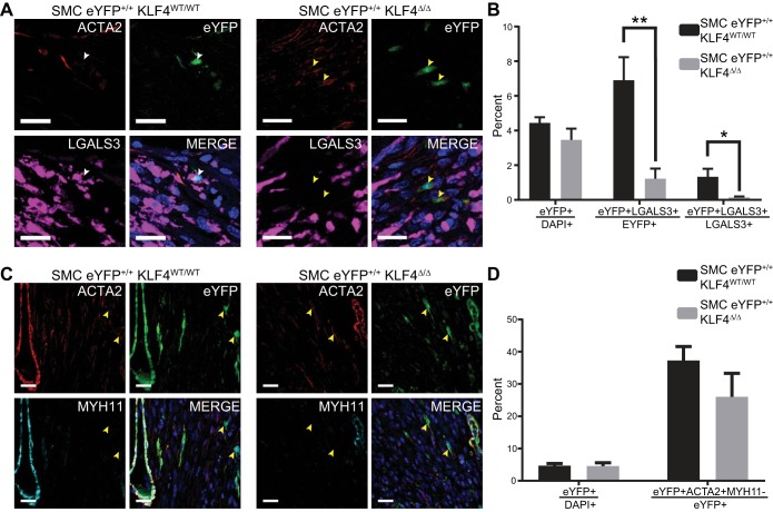
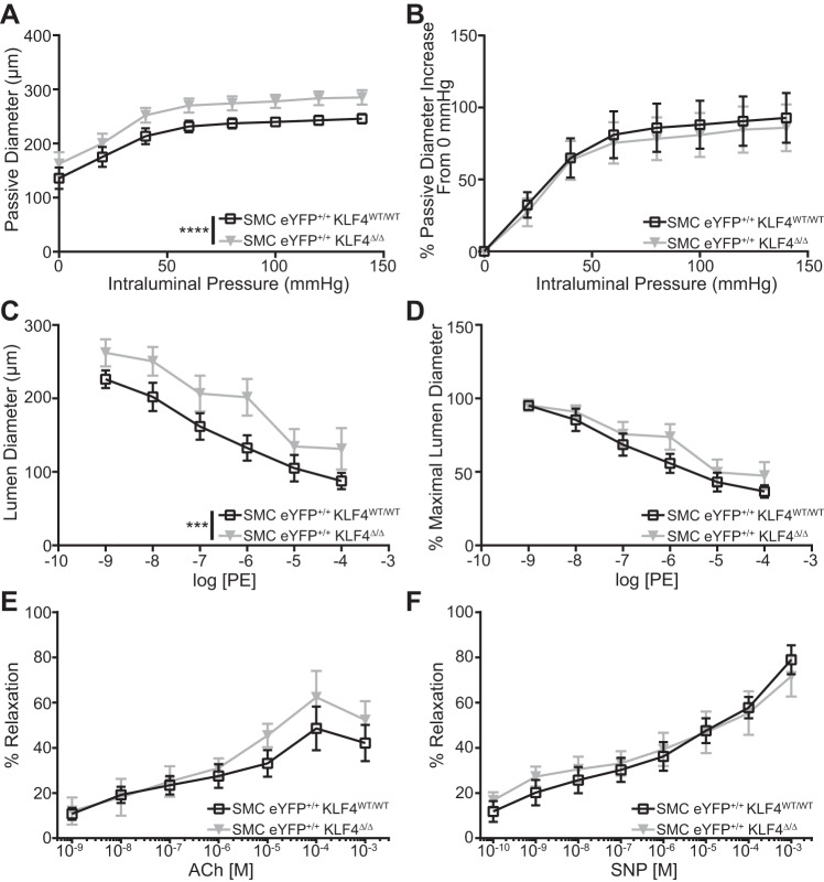
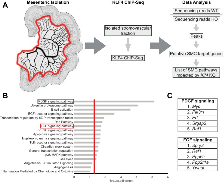
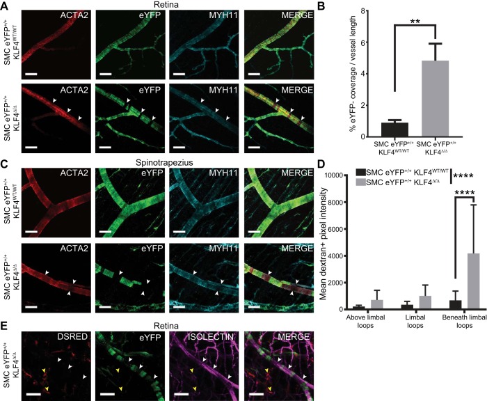
Recent smooth muscle cell (SMC) lineage-tracing studies have revealed that SMCs undergo remarkable changes in phenotype during development of atherosclerosis. Of major interest, we demonstrated that Kruppel-like factor 4 (KLF4) in SMCs is detrimental for overall lesion pathogenesis, in that SMC-specific conditional knockout of the KLF4 gene ( Klf4) resulted in smaller, more-stable lesions that exhibited marked reductions in the numbers of SMC-derived macrophage- and mesenchymal stem cell-like cells. However, since the clinical consequences of atherosclerosis typically occur well after our reproductive years, we sought to identify beneficial KLF4-dependent SMC functions that were likely to be evolutionarily conserved. We tested the hypothesis that KLF4-dependent SMC transitions play an important role in the tissue injury-repair process. Using SMC-specific lineage-tracing mice positive and negative for simultaneous SMC-specific conditional knockout of Klf4, we demonstrate that SMCs in the remodeling heart after ischemia-reperfusion injury (IRI) express KLF4 and transition to a KLF4-dependent macrophage-like state and a KLF4-independent myofibroblast-like state. Moreover, heart failure after IRI was exacerbated in SMC Klf4 knockout mice. Surprisingly, we observed a significant cardiac dilation in SMC Klf4 knockout mice before IRI as well as a reduction in peripheral resistance. KLF4 chromatin immunoprecipitation-sequencing analysis on mesenteric vascular beds identified potential baseline SMC KLF4 target genes in numerous pathways, including PDGF and FGF. Moreover, microvascular tissue beds in SMC Klf4 knockout mice had gaps in lineage-traced SMC coverage along the resistance arteries and exhibited increased permeability. Together, these results provide novel evidence that Klf4 has a critical maintenance role within microvascular SMCs: it is required for normal SMC function and coverage of resistance arteries. NEW & NOTEWORTHY We report novel evidence that the Kruppel-like factor 4 gene ( Klf4) has a critical maintenance role within microvascular smooth muscle cells (SMCs). SMC-specific Klf4 knockout at baseline resulted in a loss of lineage-traced SMC coverage of resistance arteries, dilation of resistance arteries, increased blood flow, and cardiac dilation.

Keywords: Kruppel-like factor 4; lineage tracing; smooth muscle cell maintenance; smooth muscle cells.


Figures

Fig. 1.
Fig. 1.
Kruppel-like factor 4 (KLF4) is upregulated in smooth muscle cells (SMCs) in the infarct zone after ischemia-reperfusion injury (IRI)-induced myocardial infarction (MI). A: representative immunofluorescence images of hearts from SMC eYFP+/+ mice (n = 8) at baseline. Yellow arrows highlight eYFP+KLF4 cells; white arrows highlight eYFPKlf4+ cells. ACTA2, α-smooth muscle actin. Scale bars = 20 µm. B: representative 3,3′-diaminobenzidine staining for KLF4 within hearts at baseline and 7 days post-IRI-MI. C: representative immunofluorescence images from SMC eYFP+/+Klf4WT/WT (n = 7) and SMC eYFP+/+Klf4Δ/Δ (n = 9) animals 7 days post-IRI-MI. Note marked decrease in eYFP+KLF4+ cells in SMC eYFP+/+Klf4Δ/Δ animals. White arrows highlight eYFP+KLF4+ cells; yellow arrows highlight eYFP+KLF4 cells. Scale bars = 20 µm. D: quantification of eYFP+KLF4+ cells in SMC eYFP+/+Klf4WT/WT (n = 7) and SMC eYFP+/+Klf4Δ/Δ (n = 9) animals 7 days post-IRI-MI. **P < 0.01 (by unpaired, 2-tailed t-test with Welch’s correction).
Fig. 2.
Fig. 2.
Smooth muscle cell (SMC)-specific Kruppel-like factor 4 (Klf4) knockout impairs SMC phenotypic switching to macrophage-, but not myofibroblast-like, cells. A: representative immunofluorescence images of SMC eYFP+/+Klf4WT/WT (n = 7) and SMC eYFP+/+Klf4Δ/Δ (n = 9) animals 7 days after ischemia-reperfusion injury-induced myocardial infarction (IRI-MI). Note marked decrease in eYFP+LGALS3+ cells in SMC eYFP+/+Klf4Δ/Δ animals. White arrows highlight eYFP+LGALS3+ cells; yellow arrows highlight eYFP+LGALS3 cells. ACTA2, α-smooth muscle actin; LGALS3, galectin 3. Scale bars = 20 µm. B: percent eYFP+ cells within the infarct zone, percent eYFP+ cells that are LGALS3+ within the infarct zone, and percent LGALS3+ cells that are eYFP+. Knockout of Klf4 resulted in a decrease of eYFP+LGALS3+ cells. *P < 0.05, **P < 0.01 (by unpaired, 2-tailed t-test for percent eYFP+ cells and by unpaired, 2-tailed t-test with Welch’s correction for percent eYFP+ cells that are LGALS3+ and percent LGALS3+ cells that are eYFP+). C: representative immunofluorescence images of SMC eYFP+/+Klf4WT/WT (n = 7) and SMC eYFP+/+Klf4Δ/Δ (n = 7) animals 7 days post-IRI-MI. Note no difference in eYFP+ACTA2+MYH11 cells in SMC eYFP+/+Klf4Δ/Δ compared with SMC eYFP+/+Klf4WT/WT animals. Yellow arrows highlight eYFP+ACTA2+MYH11 cells. MYH11, myosin heavy chain 11. Scale bars = 20 µm. D: quantification of percent eYFP+ cells within the infarct zone and percent eYFP+ cells that are eYFP+ACTA2+MYH11 myofibroblast-like cells within the infarct zone. *P < 0.05, **P < 0.01 (by unpaired, 2-tailed t-test).
Fig. 3.
Fig. 3.
Smooth muscle cell (SMC)-specific Kruppel-like factor 4 (Klf4) knockout leads to increased cardiac dilation after myocardial infarction (MI). A: representative images of hematoxylin-eosin staining of SMC eYFP+/+Klf4WT/WT (n = 7) and SMC eYFP+/+Klf4Δ/Δ (n = 9) animals 7 days after ischemia-reperfusion injury (IRI)-induced MI (IRI-MI). Note cardiac dilation in knockout animals. Scale bars = 1 mm. B: heart weight-to-body weight ratios for SMC eYFP+/+Klf4WT/WT (n = 3) and SMC eYFP+/+Klf4Δ/Δ (n = 4) animals 21 days post-IRI-MI. *P < 0.05 (by unpaired, 2-tailed t-test). C: quantification of infarct area as a ratio of left ventricle area for SMC eYFP+/+Klf4WT/WT (n = 7) and SMC eYFP+/+Klf4Δ/Δ (n = 9) animals 7 days post-IRI-MI. D and E: echocardiography time course of SMC eYFP+/+Klf4WT/WT animals at 0 (n = 12), 7 (n = 6), 14 (n = 3), and 21 (n = 2) days post-MI and SMC eYFP+/+Klf4Δ/Δ animals at 0 (n = 14), 7 (n = 9), 14 (n = 5), and 21 (n = 5) days post-MI. End-systolic and end-diastolic volumes and cardiac dilation were increased in knockout animals. **P < 0.01 (by 2-way ANOVA). #P < 0.05, ##P < 0.01 (by unpaired, 2-tailed t-test). There was also a significant increase in end-systolic and end-diastolic volume at baseline. F–I: echocardiography time courses examining stroke volume, ejection fraction, fractional shortening, and cardiac output. Note decreases, as expected, post-IRI-MI but no difference between knockout and wild-type animals.
Fig. 4.
Fig. 4.
Smooth muscle cell (SMC)-specific knockout of Kruppel-like factor 4 (Klf4) leads to left ventricular dilation, decreased blood pressure, and increased blood flow at baseline. A–C: echocardiography analysis of end-systolic volume, end-diastolic volume, and stroke volume at baseline in SMC eYFP+/+Klf4WT/WT (n = 12) and SMC eYFP+/+Klf4Δ/Δ (n = 14) animals. D: CD31+ pixel density within the left ventricle of SMC eYFP+/+Klf4WT/WT (n = 6) and SMC eYFP+/+Klf4Δ/Δ (n = 6) animals at baseline. E: blood pressure measured using a carotid blood pressure probe shows an ~5-mmHg drop in blood pressure in SMC eYFP+/+Klf4WT/WT (n = 7) and SMC eYFP+/+Klf4Δ/Δ (n = 5) animals at baseline. F: relative volumetric blood flow rate measured by contrast ultrasound in an area of interest near the femoral artery in SMC eYFP+/+Klf4WT/WT (n = 7) and SMC eYFP+/+Klf4Δ/Δ (n = 5) animals. *P < 0.05, **P < 0.01 (by unpaired, 2-tailed t-test).
Fig. 5.
Fig. 5.
First-order mesenteric resistance arteries from smooth muscle cell (SMC)-specific Kruppel-like factor 4 (Klf4) knockout animals are dilated compared with those from their wild-type counterparts. A: pressure myography assessment of passive properties of 1st-order mesenteric vessels from SMC eYFP+/+Klf4WT/WT (n = 3) and SMC eYFP+/+Klf4Δ/Δ (n = 3) animals (2 vessels per animal) at baseline. SMC eYFP+/+Klf4Δ/Δ vessels were significantly dilated compared with SMC eYFP+/+Klf4WT/WT vessels under all pressures. B: passive diameters normalized to respective diameters at 0 mmHg. C: pressure myography assessment of phenylephrine (PE) response of 1st-order mesenteric vessels from SMC eYFP+/+Klf4WT/WT (n = 3) and SMC eYFP+/+Klf4Δ/Δ (n = 3) animals at baseline (2 vessels per animal). Vessels from SMC eYFP+/+Klf4Δ/Δ animals were significantly dilated compared with those from SMC eYFP+/+Klf4WT/WT animals at all PE concentrations. D: PE-stimulated luminal diameter normalized to respective maximal luminal diameter. E: pressure myography assessment of endothelial-dependent acetylcholine (ACh) response of 1st-order mesenteric vessels from SMC eYFP+/+Klf4WT/WT (n = 3, total vessels = 4) and SMC eYFP+/+Klf4Δ/Δ (n = 3, total vessels = 3) animals at baseline. F: pressure myography assessment of endothelial-independent sodium nitroprusside (SNP) response of 1st-order mesenteric vessels from SMC eYFP+/+Klf4WT/WT (n = 3, total vessels = 7) and SMC eYFP+/+Klf4Δ/Δ (n = 3, total vessels = 8) animals at baseline. ***P < 0.001, ****P < 0.0001 (by 2-way ANOVA).
Fig. 6.
Fig. 6.
Kruppel-like factor 4 (KLF4) chromatin immunoprecipitation-sequencing (ChIP-Seq) identified the PDGF signaling pathway as differentially regulated between smooth muscle cell (SMC) Klf4 knockout (KO) and wild-type (WT) animals. A: cartoon representation of the area taken for the KLF4 ChIP-Seq and analysis work flow. Red line on mesentery indicates where the tissue was cut. B: Enriched Protein Analysis Through Evolutionary Relationships (PANTHER) pathways in SMC eYFP+/+Klf4WT/WT (n = 7) compared with SMC eYFP+/+Klf4Δ/Δ (n = 7) animals. Vertical red line indicates P = 0.05 [calculated using Database for Annotation Bioinformatics Microarray Analysis (DAVID) software]. C: top-5 enriched genes in PDGF and FGF signaling pathways.
Fig. 7.
Fig. 7.
Smooth muscle cell (SMC)-specific knockout of Kruppel-like factor 4 (Klf4) results in noncontinuous lineage-traced SMC coverage along vessels in the microvasculature. A: representative whole-mount immunofluorescence images of retinas from SMC eYFP+/+Klf4WT/WT (n = 8) and SMC eYFP+/+Klf4Δ/Δ (n = 8) animals. White arrows highlight gaps in eYFP+ cell coverage that have been filled with cells expressing α-smooth muscle actin (ACTA2) and myosin heavy chain 11 (MYH11) but not eYFP. Scale bars = 50 µm. B: percent eYFP length compared with whole vessel length in SMC eYFP+/+Klf4WT/WT (n = 5) and SMC eYFP+/+Klf4Δ/Δ (n = 6) retinas. **P < 0.01 (by Mann-Whitney U-test). C: representative whole-mount immunofluorescence images of spinotrapezius muscle from SMC eYFP+/+Klf4WT/WT (n = 8) and SMC eYFP+/+Klf4Δ/Δ (n = 8) animals. White arrows highlight gaps in eYFP+ cell coverage that have been filled with cells expressing ACTA2 and MYH11 but not eYFP. Scale bars = 50 µm. D: vascular leakage within the limbal vascular bed of the cornea based on intravital microscopic evaluation of interstitial levels of 70-kDa dextran at 0–1 h postinjection. Results show area under the curve for dextran+ pixels in the interstitial space near the vasculature in SMC eYFP+/+Klf4WT/WT (n = 7) and SMC eYFP+/+Klf4Δ/Δ (n = 7) animals. ****P < 0.0001 (by 2-way ANOVA for comparisons across the entire vascular network and Sidak’s multiple comparisons between individual locations). E: lethally irradiated SMC eYFP+/+Klf4Δ/Δ animals received a bone marrow transfer from whole animal constitutive DsRED+ mice. No DsRED+ cells (yellow arrows) were found in the eYFP+ SMC gaps in the spinotrapezius muscle (white arrows). Scale bars = 50 µm.

References

    1. Alexander MR, Owens GK. Epigenetic control of smooth muscle cell differentiation and phenotypic switching in vascular development and disease. Annu Rev Physiol 74: 13–40, 2012. doi:10.1146/annurev-physiol-012110-142315. - DOI - PubMed
    1. Andrae J, Gallini R, Betsholtz C. Role of platelet-derived growth factors in physiology and medicine. Genes Dev 22: 1276–1312, 2008. doi:10.1101/gad.1653708. - DOI - PMC - PubMed
    1. Armulik A, Genové G, Betsholtz C. Pericytes: developmental, physiological, and pathological perspectives, problems, and promises. Dev Cell 21: 193–215, 2011. doi:10.1016/j.devcel.2011.07.001. - DOI - PubMed
    1. Billaud M, Lohman AW, Straub AC, Parpaite T, Johnstone SR, Isakson BE. Characterization of the thoracodorsal artery: morphology and reactivity. Microcirculation 19: 360–372, 2012. doi:10.1111/j.1549-8719.2012.00172.x. - DOI - PMC - PubMed
    1. Chen PY, Qin L, Baeyens N, Li G, Afolabi T, Budatha M, Tellides G, Schwartz MA, Simons M. Endothelial-to-mesenchymal transition drives atherosclerosis progression. J Clin Invest 125: 4514–4528, 2015. doi:10.1172/JCI82719. - DOI - PMC - PubMed

Publication types

MeSH terms

Substances