

Clostridium perfringens panophthalmitis and orbital cellulitis: a case report
- PMID: 29631556
- PMCID: PMC5892009
- DOI: 10.1186/s12886-018-0751-0
Clostridium perfringens panophthalmitis and orbital cellulitis: a case report
Abstract
Background: Clostridium perfringens is an uncommon pathogen in endophthalmitis, causing rapid destruction of ocular tissues. Clostridium perfringens infection typically occurs after penetrating injury with soil-contaminated foreign bodies.
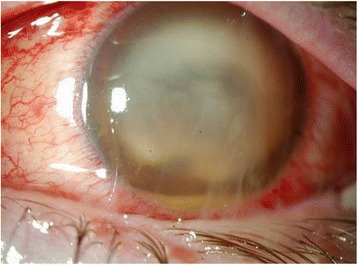
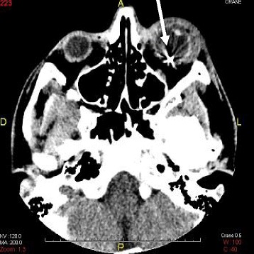
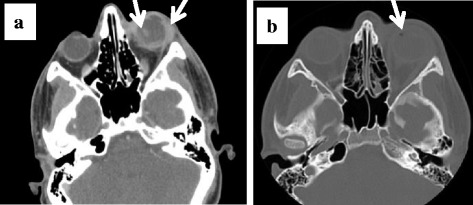
Case report: Here, we describe the case of a 17-year-old male who sustained a penetrating injury with a metallic intraocular foreign body and who rapidly developed severe C. perfringens panophthalmitis with orbital cellulitis. He was managed by systemic and intravitreal antibiotics, resulting in preservation of the globe, but a poor visual outcome.
Conclusion: Clostridial endophthalmitis secondary to penetrating injuries is a fulminant infection, almost always resulting in loss of the globe in the case of advanced infection. When feasible, early vitrectomy and intravitreal antibiotics should be considered in patients with penetrating eye injuries with contaminated foreign bodies.
Keywords: C. perfringens; Diagnosis; Panophthalmitis; Treatment.
Conflict of interest statement
Ethics approval and consent to participate
Not applicable.
Consent for publication
Written informed consent was obtained from the patient for publication of this case report. A copy of the written consent is available for review by the Editor-in-Chief of this journal.
Competing interests
The authors declare that they have no competing interest.
Publisher’s Note
Springer Nature remains neutral with regard to jurisdictional claims in published maps and institutional affiliations.
Figures

References
Publication types
MeSH terms
LinkOut - more resources
Full Text Sources
Other Literature Sources
