Review
. 2018 Jun;125(6):670-681.
doi: 10.1016/j.oooo.2018.02.020. Epub 2018 Mar 9.

Noninvasive diagnostic adjuncts for the evaluation of potentially premalignant oral epithelial lesions: current limitations and future directions

Affiliations
Review

Noninvasive diagnostic adjuncts for the evaluation of potentially premalignant oral epithelial lesions: current limitations and future directions

Eric C Yang et al. Oral Surg Oral Med Oral Pathol Oral Radiol. 2018 Jun.

Abstract

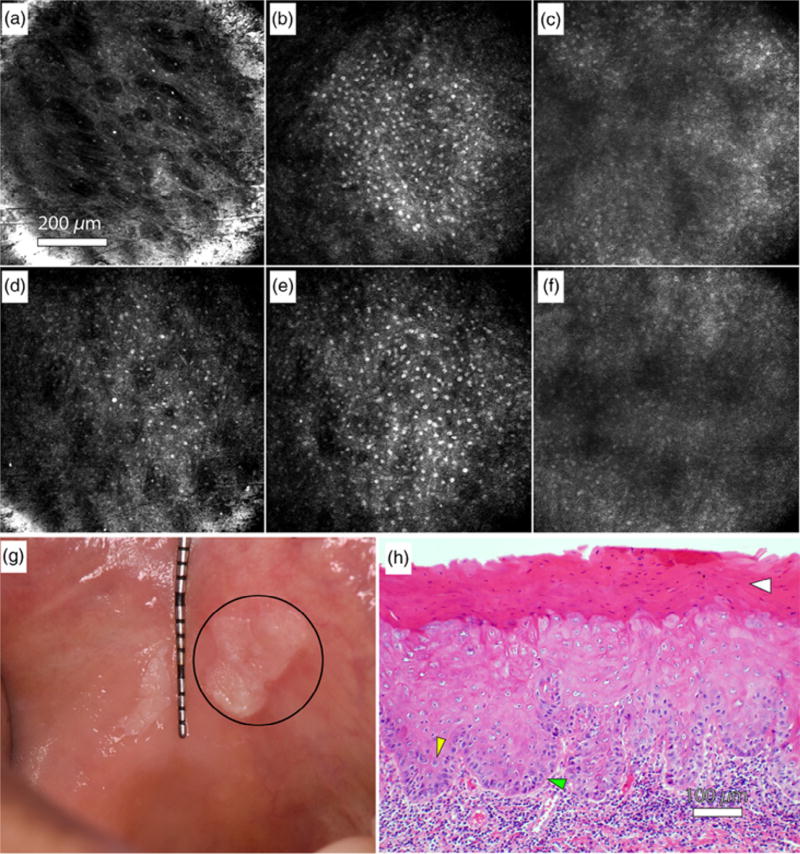
Potentially premalignant oral epithelial lesions (PPOELs) are a group of clinically suspicious conditions, of which a small percentage will undergo malignant transformation. PPOELs are suboptimally diagnosed and managed under the current standard of care. Dysplasia is the most well-established marker to distinguish high-risk PPOELs from low-risk PPOELs, and performing a biopsy to establish dysplasia is the diagnostic gold standard. However, a biopsy is limited by morbidity, resource requirements, and the potential for underdiagnosis. Diagnostic adjuncts may help clinicians better evaluate PPOELs before definitive biopsy, but existing adjuncts, such as toluidine blue, acetowhitening, and autofluorescence imaging, have poor accuracy and are not generally recommended. Recently, in vivo microscopy technologies, such as high-resolution microendoscopy, optical coherence tomography, reflectance confocal microscopy, and multiphoton imaging, have shown promise for improving PPOEL patient care. These technologies allow clinicians to visualize many of the same microscopic features used for histopathologic assessment at the point of care.


Figures

Fig. 1
Fig. 1
High-resolution microendoscopy. A, Photo of the high-resolution microendoscope (HRME) with attached probe. HRME images of histopathologically diagnosed (B) normal oral mucosa showing small, evenly spaced nuclei and (C) severe dysplasia showing enlarged, crowded nuclei. Field of view is 0.79 mm in diameter.
Fig. 2
Fig. 2
Classification of imaged sites based on autofluorescence imaging and high-resolution microendoscope (HRME). The dashed line represents a linear threshold to distinguish normal sites from moderate dysplasia to cancer sites based on imaging metrics. (Reprinted from Pierce et al.).
Fig. 3
Fig. 3
Multimodal imaging example. White light (A) and autofluorescence image (B) of red and white ulcerated lesion on the right lateral tongue in a 75-year-old female presenting at the University of Texas School of Dentistry. Site imaged with HRME (red arrow) showed loss of fluorescence with elevated normalized red-green ratio. (C) HRME probe on imaged site. (D) HRME image at site showed enlarged, crowded nuclei. Biopsy of the site was performed, and pathologic diagnosis was invasive OSCC.
Fig. 4
Fig. 4
Risk heat map based on autofluorescence imaging. White light (A) and autofluorescence image (B) of floor of mouth lesion containing histopathologically confirmed dysplasia and carcinoma in situ. C, Risk heat map based on red-green ratio overlaid on white light image, with pathologic diagnoses at 6 sites denoted by lowercase letters (a–f). Histology slides of the six sites are shown below (scale bar = 1 mm). (Reprinted from Roblyer et al.).
Fig. 5
Fig. 5
Optical coherence tomography (OCT) in the oral mucosa. A–C, Photograph (A), in vivo OCT image (B), and ×10 hematoxylin and eosin (H&E slide) (C) of dysplastic buccal mucosa. D, In vivo OCT image of normal buccal mucosa: (1) stratified squamous epithelium, (2) keratinized epithelial surface layer, (3) basement membrane, (4) submucosa. (Reprinted with permission from Wilder-Smith et al.).
Fig. 6
Fig. 6
Reflectance confocal microscopy in the oral mucosa. A–C, In vivo reflectance confocal microscopy images of clinically normal buccal mucosa at depths of approximately (A) 10 µm (superficial epithelium), (B) 70 µm (spinous layer), and (C) 120 µm (approaching basement membrane). D–F, In vivo reflectance confocal microscopy images of a leukoplakia at depths of approximately (D) 10 µm (superficial epithelium), (E) 70 µm (spinous layer), and (F) 120 µm (approaching basement membrane). G, Photograph of the leukoplakia, including the imaged lesion (circle). H, Hematoxylin and eosin (H&E) slide of imaged leukoplakia diagnosed as mild to focally moderate dysplasia. Yellow arrow: Increased nuclear to cytoplasmic ratio. Green arrow: Loss of polarity of basal cells. White arrow: Hyperkeratosis. (Reprinted from Olsovsky et al.).

References

    1. Ferlay J, Soerjomataram I, Dikshit R, et al. Cancer incidence and mortality worldwide: sources, methods and major patterns in GLOBOCAN 2012. Int J Cancer. 2015;136:E359–E386. - PubMed
    1. Warnakulasuriya S. Global epidemiology of oral and oropharyngeal cancer. Oral Oncol. 2009;45:309–316. - PubMed
    1. Warnakulasuriya S, Johnson NW, Van Der Waal I. Nomenclature and classification of potentially malignant disorders of the oral mucosa. J Oral Pathol Med. 2007;36:575–580. - PubMed
    1. Warnakulasuriya S, Ariyawardana A. Malignant transformation of oral leukoplakia: a systematic review of observational studies. J Oral Pathol Med. 2016;45:155–166. - PubMed
    1. Bouquot JE, Speight PM, Farthing PM. Epithelial dysplasia of the oral mucosa—diagnostic problems and prognostic features. Curr Diagn Pathol. 2006;12:11–21.

Publication types

MeSH terms