. 2018 May 25;62(6):e00119-18.
doi: 10.1128/AAC.00119-18. Print 2018 Jun.

High Levels of Intrinsic Tetracycline Resistance in Mycobacterium abscessus Are Conferred by a Tetracycline-Modifying Monooxygenase

Affiliations

High Levels of Intrinsic Tetracycline Resistance in Mycobacterium abscessus Are Conferred by a Tetracycline-Modifying Monooxygenase

Paulami Rudra et al. Antimicrob Agents Chemother. .

Abstract

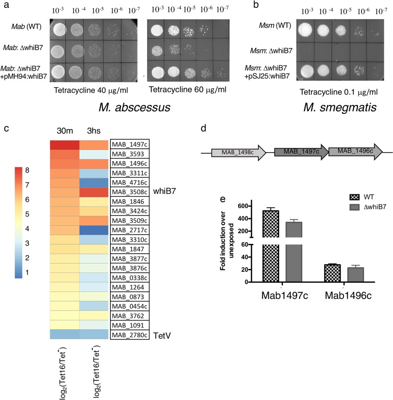
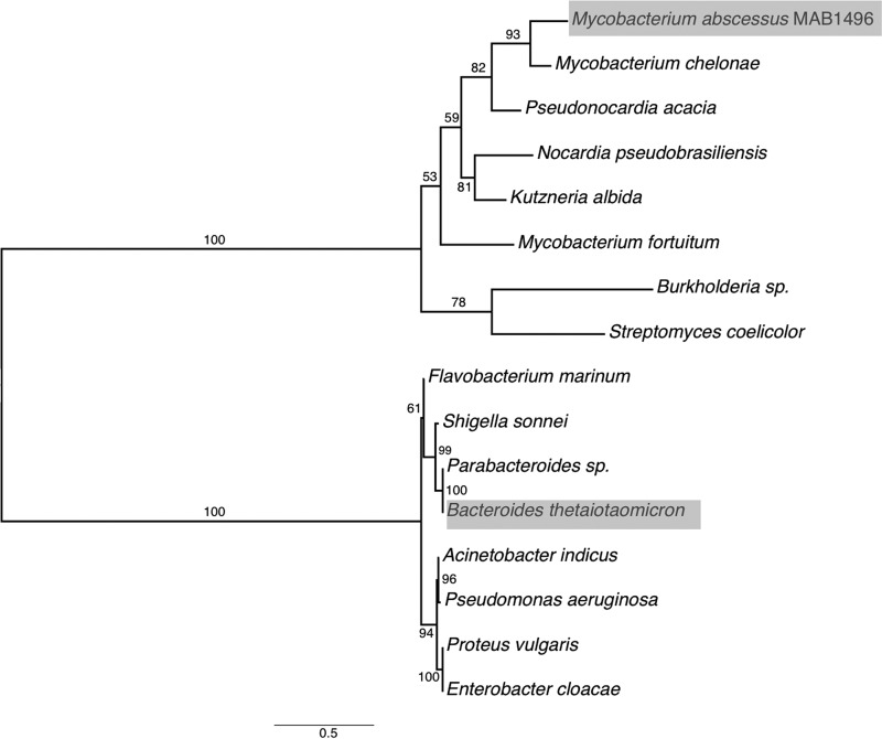
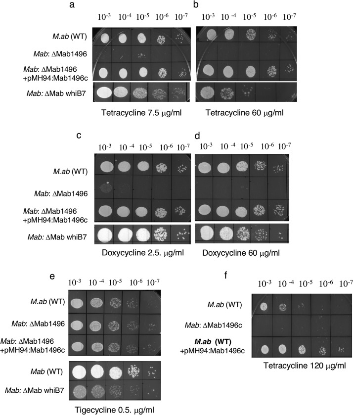
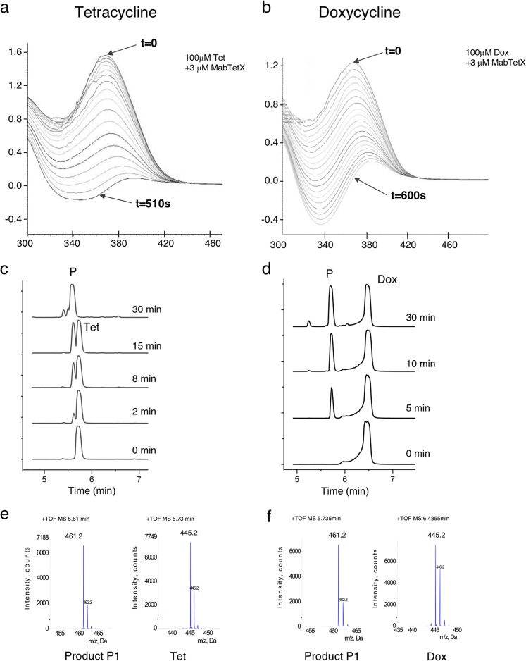
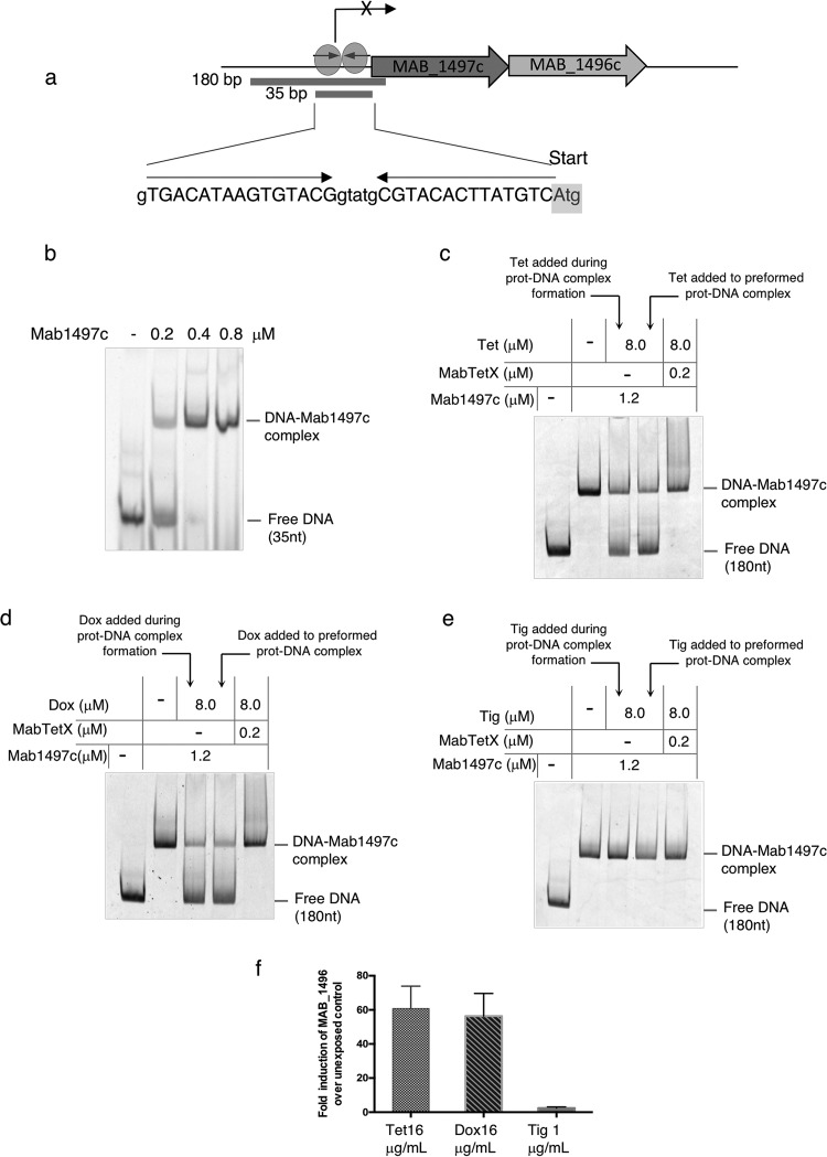
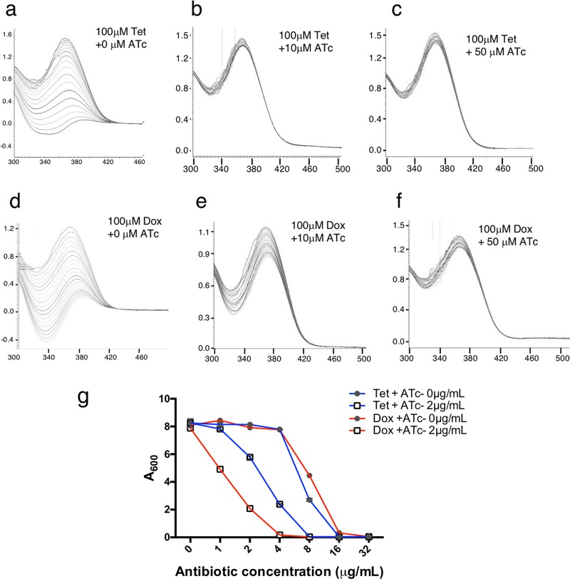
Tetracyclines have been one of the most successful classes of antibiotics. However, its extensive use has led to the emergence of widespread drug resistance, resulting in discontinuation of use against several bacterial infections. Prominent resistance mechanisms include drug efflux and the use of ribosome protection proteins. Infrequently, tetracyclines can be inactivated by the TetX class of enzymes, also referred to as tetracycline destructases. Low levels of tolerance to tetracycline in Mycobacterium smegmatis and Mycobacterium tuberculosis have been previously attributed to the WhiB7-dependent TetV/Tap efflux pump. However, Mycobacterium abscessus is ∼500-fold more resistant to tetracycline than M. smegmatis and M. tuberculosis In this report, we show that this high level of resistance to tetracycline and doxycycline in M. abscessus is conferred by a WhiB7-independent tetracycline-inactivating monooxygenase, MabTetX (MAB_1496c). The presence of sublethal doses of tetracycline and doxycycline results in a >200-fold induction of MabTetX, and an isogenic deletion strain is highly sensitive to both antibiotics. Further, purified MabTetX can rapidly monooxygenate both antibiotics. We also demonstrate that expression of MabTetX is repressed by MabTetRx, by binding to an inverted repeat sequence upstream of MabTetRx; the presence of either antibiotic relieves this repression. Moreover, anhydrotetracycline (ATc) can effectively inhibit MabTetX activity in vitro and decreases the MICs of both tetracycline and doxycycline in vivo Finally, we show that tigecycline, a glycylcycline tetracycline, not only is a poor substrate of MabTetX but also is incapable of inducing the expression of MabTetX. This is therefore the first demonstration of a tetracycline-inactivating enzyme in mycobacteria. It (i) elucidates the mechanism of tetracycline resistance in M. abscessus, (ii) demonstrates the use of an inhibitor that can potentially reclaim the use of tetracycline and doxycycline, and (iii) identifies two sequential bottlenecks-MabTetX and MabTetRx-for acquiring resistance to tigecycline, thereby reiterating its use against M. abscessus.

Keywords: M. abscessus; antibiotic resistance; drug inactivation; intrinsic resistance; tetracyclines.


Figures

FIG 1
FIG 1
Identification of MAB_1496c as a possible whiB7-independent mechanism of tetracycline resistance in M. abscessus. (a) Ten-fold serial dilutions of wild-type (WT) M. abscessus ATCC 19977, the ΔwhiB7 mutant, and the complemented strain were grown to an A600 of 0.7 and spotted onto Middlebrook 7H10 OADC containing 40 μg/ml and 60 μg/ml of tetracycline. (b) Ten-fold serial dilutions of M. smegmatis mc2155, the ΔwhiB7 mutant, and the complemented strain were grown to an A600 of 0.7 and spotted onto Middlebrook 7H10 ADC containing 0.1 μg/ml of tetracycline. (c) Transcriptomic changes in wild-type M. abscessus upon exposure to 16 μg/ml of tetracycline for 30 min and 3 h. Genes induced >4-fold with a q value < 0.001 were analyzed, and the 20 most induced genes are represented as a heat map. Expression levels of the MAB_2780c, the TetV homologue, are also included in the heat map. MAB_1496c and MAB_1497c are the 2 most highly induced genes upon tetracycline exposure. (d) Genome organization of MAB_1496c and MAB_1497c. (e) Wild-type M. abscessus and the ΔwhiB7 mutant were grown to an A600 of 0.7 and exposed to sublethal concentrations of tetracycline, and the amounts of MAB_1496c and MAB_1497c transcripts were determined by qPCR and plotted as fold induction over an unexposed control. Data represent means ± SDs (n = 3). Transcription of both genes is independent of WhiB7.
FIG 2
FIG 2
Maximum likelihood tree of MAB_1496c and Bacteroides TetX homologues. The phylogenetic tree was calculated with Phyml version 3. Numbers above each node show percent support of the branch configurations that occurred over 100 bootstrap trials. The scale bar shows the average number of amino acid residue changes per site.
FIG 3
FIG 3
Deletion of MAB_1496c renders M. abscessus hypersensitive to tetracyclines. Ten-fold serial dilutions of M. abscessus ATCC 19977, the ΔwhiB7 mutant, and the ΔMabTetX mutant were grown to an A600 of 0.7 and spotted onto Middlebrook 7H10 OADC containing the indicated concentrations of antibiotics. (a to d) The mutant is hypersensitive to tetracycline and doxycycline compared to the wild-type parent strain. A complementing strain containing an integrated copy of MAB_1496c expressed from the constitutive promoter Phsp60 restores antibiotic resistance. The sensitivity of the ΔwhiB7 mutant to tetracycline and doxycycline is apparent only at high antibiotic concentrations. (e) Tigecycline sensitivity of the ΔMAB_1496c mutant is indistinguishable from that of the wild-type strain, but the ΔwhiB7 mutant is more sensitive to tigecycline than the ΔMabTetX strain. (f) Overexpression of MAB_1496c in wild-type M. abscessus increases the resistance above that of the wild-type parent.
FIG 4
FIG 4
MAB_1496c-mediated modification of tetracycline and doxycycline. (a and b) UV-visible spectra of tetracycline and doxycycline modification catalyzed by MAB_1496c. Purified MAB_1496c was added to 100 μM tetracycline in a buffer containing 10 mM Tris (pH 8.0), 0.1 mM MgCl2, and 0.4 mM NADPH. The UV-Vis spectrum of tetracycline and doxycycline was scanned for the indicated periods with a 30-s scan interval and shows a temporal decrease in the peak at ∼370 nm. (c and d) Chromatographic separation of tetracycline and doxycycline and MAB_1496c protein-catalyzed products on an Acquity UPLC T3 (Waters, Milford, MA) column shows the appearance of an increasing product peak (P1) with time. (e and f) LC-MS was performed in a positive ion mode on a QTARXL (ABSCIEX) system. Analysis of tetracycline and doxycycline (m/z = 445.2) and their respective product peaks (m/z = 461.2) is consistent with monooxidation of each antibiotic.
FIG 5
FIG 5
MAB_1497c is a TFTR repressor that regulates the expression of MabTetX. (a) Genomic organization of MAB_1496c-MAB_ 1497c and the 35-nt palindromic sequence upstream of MAB_1497c. The 35-bp and 180-bp DNA substrates are shown. (b and e) DNA binding ability of purified MAB_1497c was analyzed using EMSA. DNA substrates were either 35 bp or 180 bp in length (as indicated) and were fluorescein labeled at the 5′ end. Addition of tetracycline and doxycycline (but not tigecycline) either during or after MAB_1497c-DNA complex formation results in disruption of DNA-protein complex. (f) Wild-type M. abscessus was grown to an A600 of 0.7 and exposed to sublethal concentrations of tetracycline, doxycycline, and tigecycline, and the amount of MAB_1496c transcripts was determined by qPCR and plotted as fold induction over an unexposed control. Data represent means ± SDs (n = 3). Tetracycline and doxycycline, but not tigecycline, induced the expression of MAB_1496c (MabTetX).
FIG 6
FIG 6
Anhydrotetracycline (ATc) is an inhibitor of MabTetX. (a to f) UV-visible spectra of tetracycline and doxycycline degradation catalyzed by MAB_1496c protein in the presence of indicated concentrations of ATc. The UV-Vis spectrum of tetracycline and doxycycline was scanned for indicated periods of time with a 30-s scan interval and shows an inhibition of the temporal decrease in the peak at ∼370 nm in the presence of ATc. (g) Wild-type M. abscessus was grown in the presence of increasing concentrations of tetracycline (blue)/doxycycline (red) in either the presence or absence of 2 mM ATc. The presence of ATc resulted in a 2- to 4-fold increase in MICs of both tetracycline and doxycycline. Data represent means ± SDs (n = 3).

Similar articles

Cited by

References

    1. Brown-Elliott BA, Wallace RJ Jr. 2002. Clinical and taxonomic status of pathogenic nonpigmented or late-pigmenting rapidly growing mycobacteria. Clin Microbiol Rev 15:716–746. doi:10.1128/CMR.15.4.716-746.2002. - DOI - PMC - PubMed
    1. Marras TK, Mendelson D, Marchand-Austin A, May K, Jamieson FB. 2013. Pulmonary nontuberculous mycobacterial disease, Ontario, Canada, 1998–2010. Emerg Infect Dis 19:1889–1891. doi:10.3201/eid1911.130737. - DOI - PMC - PubMed
    1. Lai CC, Tan CK, Chou CH, Hsu HL, Liao CH, Huang YT, Yang PC, Luh KT, Hsueh PR. 2010. Increasing incidence of nontuberculous mycobacteria, Taiwan, 2000–2008. Emerg Infect Dis 16:294–296. doi:10.3201/eid1602.090675. - DOI - PMC - PubMed
    1. Griffith DE, Aksamit T, Brown-Elliott BA, Catanzaro A, Daley C, Gordin F, Holland SM, Horsburgh R, Huitt G, Iademarco MF, Iseman M, Olivier K, Ruoss S, von Reyn CF, Wallace RJ Jr, Winthrop K, ATS Mycobacterial Diseases Subcommittee. 2007. An official ATS/IDSA statement: diagnosis, treatment, and prevention of nontuberculous mycobacterial diseases. Am J Respir Crit Care Med 175:367–416. doi:10.1164/rccm.200604-571ST. - DOI - PubMed
    1. Esther CR Jr, Esserman DA, Gilligan P, Kerr A, Noone PG. 2010. Chronic Mycobacterium abscessus infection and lung function decline in cystic fibrosis. J Cyst Fibros 9:117–123. doi:10.1016/j.jcf.2009.12.001. - DOI - PMC - PubMed

Publication types

MeSH terms

LinkOut - more resources