. 2018 May 15;200(10):3612-3625.
doi: 10.4049/jimmunol.1701287. Epub 2018 Apr 9.

Inflammatory Macrophage Expansion in Pulmonary Hypertension Depends upon Mobilization of Blood-Borne Monocytes

Affiliations

Inflammatory Macrophage Expansion in Pulmonary Hypertension Depends upon Mobilization of Blood-Borne Monocytes

Jonathan Florentin et al. J Immunol. .

Abstract

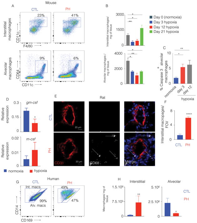
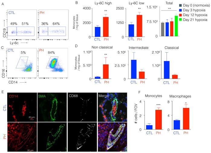
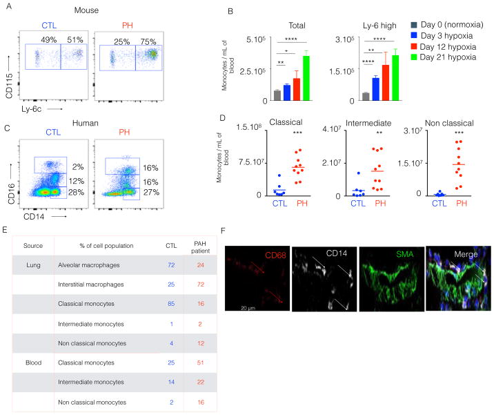
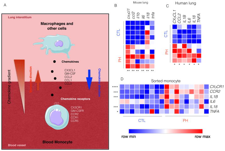
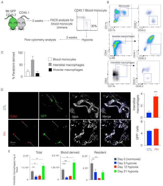
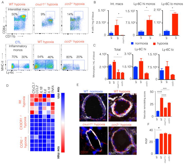
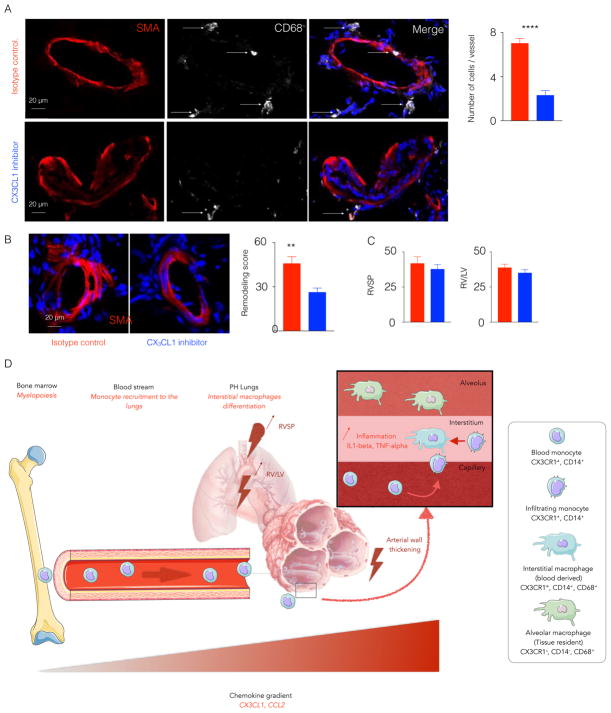
Pulmonary inflammation, which is characterized by the presence of perivascular macrophages, has been proposed as a key pathogenic driver of pulmonary hypertension (PH), a vascular disease with increasing global significance. However, the mechanisms of expansion of lung macrophages and the role of blood-borne monocytes in PH are poorly understood. Using multicolor flow cytometric analysis of blood in mouse and rat models of PH and patients with PH, an increase in blood monocytes was observed. In parallel, lung tissue displayed increased chemokine transcript expression, including those responsible for monocyte recruitment, such as Ccl2 and Cx3cl1, accompanied by an expansion of interstitial lung macrophages. These data indicate that blood monocytes are recruited to lung perivascular spaces and differentiate into inflammatory macrophages. Correspondingly, parabiosis between congenically different hypoxic mice demonstrated that most interstitial macrophages originated from blood monocytes. To define the actions of these cells in PH in vivo, we reduced blood monocyte numbers via genetic deficiency of cx3cr1 or ccr2 in chronically hypoxic male mice and by pharmacologic inhibition of Cx3cl1 in monocrotaline-exposed rats. Both models exhibited decreased inflammatory blood monocytes, as well as interstitial macrophages, leading to a substantial decrease in arteriolar remodeling but with a less robust hemodynamic effect. This study defines a direct mechanism by which interstitial macrophages expand in PH. It also demonstrates a pathway for pulmonary vascular remodeling in PH that depends upon interstitial macrophage-dependent inflammation yet is dissociated, at least in part, from hemodynamic consequences, thus offering guidance on future anti-inflammatory therapeutic strategies in this disease.


Figures

Figure 1
Figure 1. The number of lung interstitial macrophages is increased in hypoxic mice, monocrotaline-injected rats and patients
C57BL/6 mice were placed in normoxia or hypoxia (n=5 per group) for three weeks to induce PH. A) Representative flow cytometric plots for murine alveolar and interstitial macrophages in the lungs are shown, with B) quantification of these cells at different time points after hypoxia exposure. C) Percentage of caspase 3 positive alveolar macrophages in normoxic and hypoxic mice was determined by flow cytometry. D) Quantification of gm-csf and m-csf in the lungs of hypoxic mice compared to normoxic controls. E) Confocal imaging of lung sections were stained for CD31 and CD68 in monocrotaline-injected rats, and F) quantification of CD68+ interstitial perivascular macrophages in monocrotaline-injected rats was pursued. n=5 control rats and 5 MCT rats. G) Flow cytometric plots display the proportions of human alveolar vs. interstitial macrophages in control (CTL) and PH lung samples. H) Quantitation of human alveolar and interstitial macrophages is shown. n=5 healthy donors and 6 PAH patients. Mean ± s.e.m. * P < 0.05, ** P < 0.01, **** P < 0.001.
Figure 2
Figure 2. The number of inflammatory lung monocytes is elevated in hypoxic mice and PAH patients
C57BL/6 mice were placed in a 10% O2 hypoxic chamber (n=4 normoxic controls and 6 hypoxic mice) for three weeks to induce PH. A) Flow cytometric plots showing the proportion of Ly-6Chigh and Ly-6Clow monocytes in hypoxic and normoxic mouse lung samples. B) Quantitation of Ly-6Chigh and Ly-6Clow monocytes at 21 days, and total monocytes at various timepoints after hypoxia initiation C) PAH patient lung samples (n=6) or control lung samples (n=5) were collected. Flow cytometric plots showing the proportion of lung non-classical monocytes in control (CTL) and PAH patients. D) Quantitation of classical, intermediate and non-classical monocytes in CTL and PAH lung samples. E) Confocal imaging of patient lung sections stained for CD68, SMA and CD14. F) Quantification of monocytes and macrophages in CTL and PAH lung samples. Mean ± s.e.m. ** P < 0.01.
Figure 3
Figure 3. Circulatory inflammatory monocytes were increased in hypoxic mice and PAH patients
C57BL/6 mice were placed in a 10% O2 hypoxic chamber (n=4 normoxic and 6 hypoxic mice) for three weeks to induce PH. A) The flow cytometric plots show the proportion of circulatory Ly-6Chigh and Ly-6Clow monocytes in hypoxic and normoxic mice and B) Quantification of total monocytes and Ly-6Chigh monocytes at various timepoints after hypoxic exposure. C) Blood from PAH patients (n=9) and healthy donors (n=7) was collected. Flow cytometric plots show the proportion of blood inflammatory monocytes in control (CTL) and PH patients. D) Quantification of classical, intermediate and non-classical monocytes is shown in both CTL and PAH blood samples. E) Blood and lung samples were collected from a PH patient. The frequency of lung macrophage and circulatory monocyte subsets of a healthy control and the PAH patient is shown in the table. F) Confocal images of lung sections of the PH patient are displayed. The sections were stained for CD68, SMA and CD14. Mean ± s.e.m., ** P < 0.01, *** P<0.001 **** P < 0.0001.
Figure 4
Figure 4. Hypoxic mice and patients with PH had increased levels of cytokines, chemokines and chemokine receptors
C57BL/6 mice were placed in a 10% O2 hypoxic chamber (n=5 per group) for three weeks to induce PH. A) Schematic diagram shows that lung chemokines trigger the active recruitment of blood monocytes to the lungs. Adapted from the Medical Servier of Art. B) Quantitation of cx3cl1, ccl2, il1b, il6, il18 and tnfa mRNA expression in the lungs of hypoxic and normoxic mice. Blood and lung samples from PAH patients (n=9) and healthy donors (n=7) were collected. C) Quantitation of CX3CL1, CCL2, IL1B, IL6, IL18, and TNFA expression in the lungs of PH patients and healthy controls. D) Quantitation of chemokine receptors and pro-inflammatory cytokines mRNA expression such as CX3CL1, CCL2, IL1B, IL6, IL18, and TNFA by monocytes sorted from the blood of PH patients and healthy controls. Mean ± s.e.m., * P < 0.05 ** P < 0.01, *** P< 0.001 **** P < 0.0001.
Figure 5
Figure 5. Parabiosis between hypoxic mice demonstrated the monocytic origin of interstitial macrophages in the lungs
A) CD45.1 C57BL/6 mouse and CD45.2 GFP C57BL/6 mouse were surgically joined. Three weeks after the parabiosis, they were placed in a hypoxic chamber for 3 weeks. B) Flow cytometric plots show the chimera levels in blood monocytes and lung macrophages of hypoxic mice. C) The histogram shows the percentage of CD45.2+ parabiont-derived cells in blood monocytes, interstitial macrophages and alveolar macrophages in CD45.1 hypoxic mice. D) Confocal imaging of lung sections of the CD45.1+ parabionts shows GFP+ interstitial perivascular macrophages. E) Flow cytometric enumeration of total, monocyte-derived and resident interstitial macrophages in the lungs of hypoxic mice and normoxic controls. n=4 mice per group. Mean ± s.e.m, * P < 0.05 ** P < 0.01, *** P< 0.001 ****.
Figure 6
Figure 6. CCR2 and CX3CR1 deficient mice had an attenuated lung remodeling
ccr2−/− and cx3cr1−/− mice were put in hypoxic chambers for 3 weeks. n= 5 normoxic wild type (WT) mice, 5 hypoxic WT mice, 5 hypoxic cx3cr1−/− mice and 5 ccr2−/− hypoxic mice. A) Flow plots showing the proportions of interstitial macrophages and monocytes in WT, ccr2−/− and cx3cr1−/− mice. B) The histograms show the number of total monocytes, Ly-6Chigh monocytes and Ly-6Clow monocytes in the lungs. C) Histograms showing the number of interstitial macrophages, Ly-6Chigh monocytes and Ly-6Clow monocytes in the blood of WT, cx3cr1−/− and ccr2−/− mice. D) Quantitation of mRNA levels of chemokines and pro-inflammatory cytokines in the lungs. E) Confocal images of lung sections stained for CD31 and SMA. F) Histograms showing right ventricular systolic pressure (RVSP). Mean ± s.e.m., * P < 0.05, *** P< 0.001, **** P < 0.0001.
Figure 7
Figure 7. Cx3cl1 inhibitor limited the recruitment of monocytes in monocrotaline-injected rats
Adult male Sprague-Dawley rats were injected once with monocrotaline to induce PH. The rats were injected with either Cx3cl1 inhibitor or isotype antibody for 3 weeks. A) Confocal imaging of lung sections stained for CD68 and SMA. The histogram quantifies lung perivascular macrophages. B) The confocal images of lung sections stained for SMA show the lung vasculature remodeling. Remodeling score was quantified. C) The histograms show right ventricular systolic pressure and RV/LV ratio in isotype control and Cx3cl1 inhibitor-injected rats. n=5 per group, Mean ± s.e.m., ** P< 0.01, **** P < 0.001. D) The schematic diagram depicts monocyte recruitment to the lungs and expansion of interstitial macrophages in PH. CX3CR1+ CCR2+ monocytes egress from the bone marrow into the blood stream. The production of chemokines, such as CX3CL1 and CCL2, in the lungs and increased expression of the receptors of these chemokines on circulatory monocytes mediate their recruitment into the lungs in the context of PH. These newly recruited monocytes differentiate into interstitial perivascular macrophages. Adapted from the Medical Servier of Art.

Similar articles

Cited by

References

    1. Sawada H, et al. Reduced BMPR2 expression induces GM-CSF translation and macrophage recruitment in humans and mice to exacerbate pulmonary hypertension. J Exp Med. 2014;211(2):263–80. - PMC - PubMed
    1. Frid MG, et al. Hypoxia-induced pulmonary vascular remodeling requires recruitment of circulating mesenchymal precursors of a monocyte/macrophage lineage. Am J Pathol. 2006;168(2):659–69. - PMC - PubMed
    1. Hwangbo C, et al. Modulation of Endothelial BMPR2 Activity by VEGFR3 in Pulmonary Arterial Hypertension. Circulation. 2017
    1. Mathew R, et al. Hematological disorders and pulmonary hypertension. World J Cardiol. 2016;8(12):703–718. - PMC - PubMed
    1. Mouraret N, et al. Activation of lung p53 by Nutlin-3a prevents and reverses experimental pulmonary hypertension. Circulation. 2013;127(16):1664–76. - PMC - PubMed

Publication types