. 2018 May 2;38(18):4301-4315.
doi: 10.1523/JNEUROSCI.2751-17.2018. Epub 2018 Apr 9.

Matrix Metalloproteinase-Mediated Blood-Brain Barrier Dysfunction in Epilepsy

Affiliations

Matrix Metalloproteinase-Mediated Blood-Brain Barrier Dysfunction in Epilepsy

Ralf G Rempe et al. J Neurosci. .

Abstract

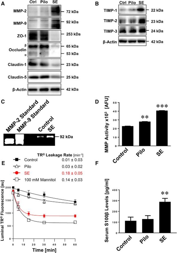
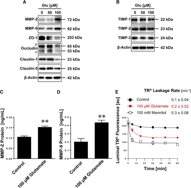
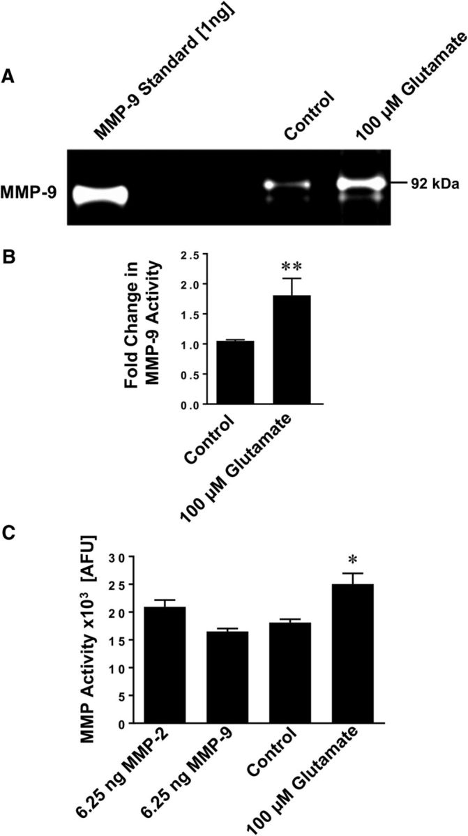
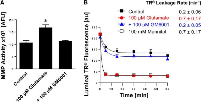
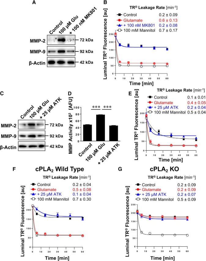
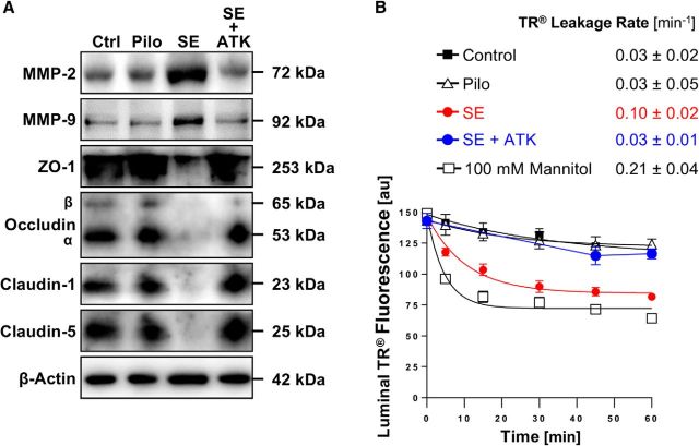
The blood-brain barrier is dysfunctional in epilepsy, thereby contributing to seizure genesis and resistance to antiseizure drugs. Previously, several groups reported that seizures increase brain glutamate levels, which leads to barrier dysfunction. One critical component of barrier dysfunction is brain capillary leakage. Based on our preliminary data, we hypothesized that glutamate released during seizures mediates an increase in matrix-metalloproteinase (MMP) expression and activity levels, thereby contributing to barrier leakage. To test this hypothesis, we exposed isolated brain capillaries from male Sprague Dawley rats to glutamate ex vivo and used an in vivo/ex vivo approach of isolated brain capillaries from female Wistar rats that experienced status epilepticus as an acute seizure model. We found that exposing isolated rat brain capillaries to glutamate increased MMP-2 and MMP-9 protein and activity levels, and decreased tight junction protein levels, which resulted in barrier leakage. We confirmed these findings in vivo in rats after status epilepticus and in brain capillaries from male mice lacking cytosolic phospholipase A2 Together, our data support the hypothesis that glutamate released during seizures signals an increase in MMP-2 and MMP-9 protein expression and activity levels, resulting in blood-brain barrier leakage.SIGNIFICANCE STATEMENT The mechanism leading to seizure-mediated blood-brain barrier dysfunction in epilepsy is poorly understood. In the present study, we focused on defining this mechanism in the brain capillary endothelium. We demonstrate that seizures trigger a pathway that involves glutamate signaling through cytosolic phospholipase A2, which increases MMP levels and decreases tight junction protein expression levels, resulting in barrier leakage. These findings may provide potential therapeutic avenues within the blood-brain barrier to limit barrier dysfunction in epilepsy and decrease seizure burden.

Keywords: Blood-Brain barrier; MMP; barrier dysfunction; barrier leakage; cPLA2.


Figures

Figure 1.
Figure 1.
Expression and activity of MMP-2 and MMP-9 in isolated brain capillaries. A, Representative immunostaining for MMP-2 (left), MMP-9 (middle), and the negative control (right; overlay of green, blue, and transmitted light channels) in isolated rat brain capillaries. Green represents MMPs. Blue represents nuclei counterstained with DAPI. B, Western blot showing MMP-2 and MMP-9 in liver lysate, liver crude membrane fraction (MF), kidney brush border membrane (BBM), brain lysate, and choroid plexus (CP) lysate. C, Western blot showing MMP-2 and MMP-9 in brain capillary (Cap) lysate and brain capillary crude membrane fraction (Cap MF). D, Total MMP enzyme activity in brain capillaries was measured using the fluorogenic substrate Mca-PLGL-Dpa-AR-NH2. MMP activity was assessed in brain capillary lysate with or without MMP inhibitor (GM6001). MMP activity is given as AFU; data are mean ± SEM (n = 3 independent experiments; pooled tissue from n = 10 rats per experiment). **Significantly lower than control (t(2) = 10.37, p = 0.0092; unpaired t test).
Figure 2.
Figure 2.
SE increases MMP levels, decreases levels of tight junction proteins, and causes barrier leakage in brain capillaries. A, Western blot showing MMP-2, MMP-9, ZO-1, occludin, claudin-1, and claudin-5 in isolated brain capillaries from rats after an SE induced with pilocarpine, rats that received pilocarpine but did not develop an SE (Pilo), and untreated control rats (Ctrl). β-Actin was used as protein loading control. B, Western blot showing TIMP-1, TIMP-2, and TIMP-3 protein expression in isolated brain capillaries from control, pilocarpine, and SE rats. C, Representative MMP-9 gelatin zymogram of brain capillary lysates from rats after SE and from control rats. Left, Positive controls from recombinant expressed MMP-2 and MMP-9. D, Total MMP enzyme activity in brain capillaries isolated from control rats, rats that received pilocarpine but did not develop an SE (Pilo), and rats after an SE was measured using the fluorogenic substrate Mca-PLGL-Dpa-AR-NH2. ***SE significantly higher than control, 1.8-fold (t(2) = 78.6, p = 0.0002; ANOVA post hoc test); Pilo significantly higher than control, 1.2-fold (t(2) = 19.0, p = 0.0028; ANOVA post hoc test); n = 2 independent experiments. E, Texas Red (TR) leakage from capillaries of rats after an SE induced by pilocarpine (SE), rats that received pilocarpine but did not develop an SE (Pilo), and untreated control rats with or without high osmotic mannitol used as positive control for barrier opening. Data are mean ± SEM for n = 7 capillaries per time point from one brain capillary isolation with n = 10 rats. Shown are 0–255 AFU. First-order efflux rates were calculated using nonlinear regression. F, S100β levels in serum samples from control rats, rats that received pilocarpine but did not develop an SE (Pilo), and rats after an SE was determined by ELISA. **Control versus SE: t(2) = 3.79, p > 0.0012; Pilo versus SE: t(2) = 3.58, p > 0.002 (ANOVA post hoc tests). n = 2 independent experiments.
Figure 3.
Figure 3.
Glutamate increases MMP-2 and MMP-9 levels, decreases tight junction protein levels, and causes barrier leakage in isolated brain capillaries. A, Western blot showing MMP-2, MMP-9, ZO-1, occludin, claudin-1, and claudin-5 in isolated rat brain capillaries exposed to 0, 50, or 100 μm glutamate (Glu); β-actin was used as protein loading control. B, Western blot showing TIMP-1, TIMP-2, and TIMP-3 protein expression in isolated brain capillaries from control, pilocarpine, and SE rats. C, MMP-2 protein levels in rat brain capillary lysate determined by ELISA from control capillaries and capillaries exposed to 100 μm glutamate. Data are mean ± SEM (n = 3 independent experiments; pooled tissue of n = 10 rats per experiment). Statistical comparison: **t(4) = 6.65, p = 0.0027 (unpaired t test). D, MMP-9 protein levels in rat brain capillary lysate determined by ELISA from control capillaries and capillaries exposed to 100 μm glutamate. Data are mean ± SEM (n = 3 independent experiments; pooled tissue of n = 10 rats per experiment). Statistical comparison: **t(4) = 4.63, p = 0.0098 (unpaired t test). E, Texas Red leakage from rat brain capillaries exposed to 100 μm glutamate; high osmotic mannitol was used as positive control for barrier opening. Data are 0–255 AFU and presented as mean ± SEM for n = 7 brain capillaries per time point from one brain capillary isolation with n = 10 rats.
Figure 4.
Figure 4.
Glutamate increases MMP activity in isolated brain capillaries. A, Representative MMP-9 gelatin zymogram of lysate from brain capillaries exposed to 100 μm glutamate. Left, Recombinant MMP-9 (positive control). B, Densitometric analysis of n = 4 gelatin zymograms. Data are mean ± SEM (n = 4 independent experiments) and show the fold change over controls (1.81 ± 0.28-fold; t(6) = 5.77, p = 0.0012; unpaired t test) for MMP-9 activity in lysate from rat brain capillaries exposed to 100 μm glutamate. C, Total MMP enzyme activity in brain capillaries was measured using the fluorogenic substrate Mca-PLGL-Dpa-AR-NH2. MMP activity was measured in lysate from isolated brain capillaries exposed to 100 μm glutamate; recombinant rat MMP-2 and rat MMP-9 were used as positive controls. MMP activity is given as AFU; data are mean ± SEM (n = 3 independent experiments). *Significantly lower than control (t(4) = 3.44, p = 0.026, unpaired t test). **Significantly higher than control, p < 0.01.
Figure 5.
Figure 5.
Effect of inhibiting MMPs on glutamate-mediated barrier leakage. A, Total MMP enzyme activity in brain capillaries was assessed by using the fluorogenic substrate Mca-PLGL-Dpa-AR-NH2. MMP activity was measured in lysate from isolated brain capillaries exposed to 100 μm glutamate with or without the MMP inhibitor GM6001. MMP activity is given as AFU; data are mean ± SEM (n = 3 independent experiments; pooled tissue of 10 rats per experiment). Statistical comparison: *control: 10.7 ± 0.6 AFU; glutamate: 16.8 ± 0.8 AFU; glutamate + GM6001: 11.3 ± 0.2 AFU; glutamate + GM6001 versus control: t(2) = 0.69, p = 0.56; glutamate + GM6001 versus glutamate: t(2) = 4.83, p = 0.04 (ANOVA post hoc tests). B, Texas Red leakage was measured in capillaries exposed to 100 μm glutamate with or without the MMP inhibitor GM6001.
Figure 6.
Figure 6.
Effect of inhibiting cPLA2 on glutamate-mediated MMP-2 and MMP-9 induction and barrier leakage. A, Western blots showing MMP-2 and MMP-9 protein expression in isolated rat brain capillaries exposed to 100 μm glutamate with or without the NMDAR antagonist MK801. β-Actin was used as protein loading control. B, Texas Red leakage assay showing glutamate-mediated barrier leakage with or without the NMDAR antagonist MK801. C, Western blots showing MMP-2 and MMP-9 protein expression levels in isolated rat brain capillaries exposed to 100 μm glutamate with or without the cPLA2 inhibitor ATK. β-Actin was used as protein loading control. D, MMP activity assessed in isolated capillaries exposed to 100 μm glutamate with or without ATK (ATK vs control: t(2) = 3.08, p = 0.091; ANOVA post hoc test). E, Texas Red leakage assay showing glutamate-mediated barrier leakage with or without ATK (cPLA2 inhibitor). Texas Red leakage assay in capillaries from (F) wild-type and (G) cPLA2 KO mice that were exposed to 100 μm glutamate with or without the cPLA2 inhibitor ATK. Data are 0–255 AFU and presented as mean ± SEM for n = 7 brain capillaries per time point from one capillary isolation with n = 10 rats or n = 30 mice, respectively. ***Significantly higher than control, p < 0.001.
Figure 7.
Figure 7.
Inhibition of cPLA2 in vivo in SE rats. A, Western blot showing MMP-2, MMP-9, ZO-1, occludin, claudin-1, and claudin-5 in brain capillaries isolated from control rats (Ctrl) and from rats that experienced pilocarpine-induced SE, that received pilocarpine but did not develop SE (Pilo), and that experienced SE and received the cPLA2 inhibitor ATK (SE + ATK). B, Texas Red leakage in brain capillaries from control rats (n = 6), pilocarpine control rats (n = 4), SE control rats (n = 6), and ATK-treated SE rats (n = 6). High osmotic mannitol was used as positive marker for capillary leakage. Data are 0–255 AFU and are presented as mean ± SEM for n = 7 brain capillaries per time point from pooled tissue per group.
Figure 8.
Figure 8.
Proposed signaling pathway.

Comment in

References

    1. Alonso-Nanclares L, DeFelipe J (2014) Alterations of the microvascular network in the sclerotic hippocampus of patients with temporal lobe epilepsy. Epilepsy Behav 38:48–52. 10.1016/j.yebeh.2013.12.009 - DOI - PubMed
    1. András IE, Deli MA, Veszelka S, Hayashi K, Hennig B, Toborek M (2007) The NMDA and AMPA/KA receptors are involved in glutamate-induced alterations of occludin expression and phosphorylation in brain endothelial cells. J Cereb Blood Flow Metab 27:1431–1443. 10.1038/sj.jcbfm.9600445 - DOI - PubMed
    1. Baik EJ, Kim EJ, Lee SH, Moon C (1999) Cyclooxygenase-2 selective inhibitors aggravate kainic acid induced seizure and neuronal cell death in the hippocampus. Brain Res 843:118–129. 10.1016/S0006-8993(99)01797-7 - DOI - PubMed
    1. Bankstahl JP, Hoffmann K, Bethmann K, Löscher W (2008) Glutamate is critically involved in seizure-induced overexpression of P-glycoprotein in the brain. Neuropharmacology 54:1006–1016. 10.1016/j.neuropharm.2008.02.008 - DOI - PubMed
    1. Barr TL, Latour LL, Lee KY, Schaewe TJ, Luby M, Chang GS, El-Zammar Z, Alam S, Hallenbeck JM, Kidwell CS, Warach S (2010) Blood-Brain barrier disruption in humans is independently associated with increased matrix metalloproteinase-9. Stroke 41:e123–128. 10.1161/STROKEAHA.109.570515 - DOI - PMC - PubMed

Publication types