. 2018 Apr 24;115(17):E4071-E4080.
doi: 10.1073/pnas.1719267115. Epub 2018 Apr 9.

Human hydroxymethylbilane synthase: Molecular dynamics of the pyrrole chain elongation identifies step-specific residues that cause AIP

Affiliations

Human hydroxymethylbilane synthase: Molecular dynamics of the pyrrole chain elongation identifies step-specific residues that cause AIP

Navneet Bung et al. Proc Natl Acad Sci U S A. .

Abstract

Hydroxymethylbilane synthase (HMBS), the third enzyme in the heme biosynthetic pathway, catalyzes the head-to-tail condensation of four molecules of porphobilinogen (PBG) to form the linear tetrapyrrole 1-hydroxymethylbilane (HMB). Mutations in human HMBS (hHMBS) cause acute intermittent porphyria (AIP), an autosomal-dominant disorder characterized by life-threatening neurovisceral attacks. Although the 3D structure of hHMBS has been reported, the mechanism of the stepwise polymerization of four PBG molecules to form HMB remains unknown. Moreover, the specific roles of each of the critical active-site residues in the stepwise enzymatic mechanism and the dynamic behavior of hHMBS during catalysis have not been investigated. Here, we report atomistic studies of HMB stepwise synthesis by using molecular dynamics (MD) simulations, mutagenesis, and in vitro expression analyses. These studies revealed that the hHMBS active-site loop movement and cofactor turn created space for the elongating pyrrole chain. Twenty-seven residues around the active site and water molecules interacted to stabilize the large, negatively charged, elongating polypyrrole. Mutagenesis of these active-site residues altered the binding site, hindered cofactor binding, decreased catalysis, impaired ligand exit, and/or destabilized the enzyme. Based on intermediate stages of chain elongation, R26 and R167 were the strongest candidates for proton transfer to deaminate the incoming PBG molecules. Unbiased random acceleration MD simulations identified R167 as a gatekeeper and facilitator of HMB egress through the space between the enzyme's domains and the active-site loop. These studies identified the specific active-site residues involved in each step of pyrrole elongation, thereby providing the molecular bases of the active-site mutations causing AIP.

Keywords: enzyme catalysis; molecular dynamics; structural biology.


Conflict of interest statement

The authors declare no conflict of interest.

Figures
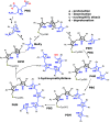
Fig. 1.
Fig. 1.
Schematic representation of mechanism of tetrapyrrole chain elongation catalyzed by hHMBS. The figure shows (a) protonation of PBG, (b) deamination of PBG to form MePy, and (c) nucleophilic attack by ring B of DPM on MePy, forming an intermediate that (d) undergoes deprotonation to form a tripyrrole moiety (P3M). Subsequent additions of PBG elongate the chain to form tetrapyrrole (P4M), pentapyrrole (P5M), and hexapyrrole (P6M) moieties. At the end of the last step, the tetrapyrrole product HMB is hydrolyzed, leaving the DPM cofactor attached to protein. The rings of the elongating pyrrole chain are labeled as A, B, C, D, E, and F starting from the pyrrole ring covalently attached to C261. The acetate and propionate side groups of the pyrroles are denoted by “-Ac” and “-Pr,” respectively.
Fig. 2.
Fig. 2.
Structure of hHMBS (PDB ID code 3ECR) with modeled missing residues showing the domains (1, 2, and 3 in blue, red, and green, respectively) with hinge regions (115–119, 213–218, and 237–240 in pink), the additional 29-residue insert (296–324 in orange), and the active-site loop (56–76 in cyan).
Fig. 3.
Fig. 3.
Structural fluctuations during polypyrrole accommodation. (A) rmsd of the protein along with domainwise rmsd, with reference to the DPM stage structure, during stages of chain elongation. (B) RMSF of Cα atoms of hHMBS protein at each stage of chain elongation, from DPM to P6M. The encircled regions show high fluctuations in the active-site loop and 29-residue insert regions. (C) The distance between the centers of mass of the active-site loop and the active-site residues (R26, Q34, D99, R149, and R150) as a function of time along the stages of chain elongation emphasizes the role of the loop in polypyrrole accommodation. The SE is shown as error bars along y-coordinates. (D) The cofactor turn shifts in the P4M stage (green) compared with the DPM stage (cyan) to accommodate the growing pyrrole chain during the stages of chain elongation. (E) Characterization of the active-site loop conformation as a function of (i) distance between the centers of mass of the active-site residues (R26, Q34, D99, R149, and R150) and the active-site loop on the x-axis and (ii) the rmsd of the active-site loop on the y-axis along the concatenated trajectory and (F) the conformation of the active-site loop at bins 1, 2, and 3 shown in red, blue, and green, respectively. DPM is shown in yellow and P6M is shown in green sticks.
Fig. 4.
Fig. 4.
Residues in active site stabilize growing polypyrrole chain. Interactions of the residues lining the active site with the growing pyrrole chain at (A) DPM, (B) P3M, (C) P4M, (D) P5M, and (E) P6M stages. The interactions are shown by dotted lines. The cofactor is shown in yellow, and water molecules are represented as spheres. The carboxylate oxygen (marked as “O”) of acetate/propionate side chains and the pyrrole nitrogen (marked as “N”) for each of the pyrrole rings are labeled. The numeric suffixes (labeled 1–4) indicate the positions of the oxygen on the pyrrole, and the letter suffixes (“A” through “F”) indicate the respective pyrrole rings from which the carboxylate oxygen is attached.
Fig. 5.
Fig. 5.
Water-mediated interactions stabilize the negative charge on polypyrrole. Graph showing the total number of water molecules interacting with the polypyrrole during the stages of chain elongation (black); total number of water-mediated interactions between the growing polypyrrole and protein (red).
Fig. 6.
Fig. 6.
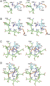
R26, in DPM and P3M stages, and R167, in P4M and P5M stages, mediate the catalytic mechanism during substrate addition. Stereograms of PBG docked in the active site of hHMBS at (A) DPM, (B) P3M, (C) P4M, and (D) P5M stages showcase the role of R26 and R167 in proton donation to the incoming PBG. All measurements are in angstroms. The PBG, polypyrrole, arginine (R26 and R167), and aspartate (D99) residues are shown in cyan, green, orange, and violet colors, respectively.
Fig. 7.
Fig. 7.
Exit of HMB from the catalytic cleft of hHMBS. (A) Three possible exit paths for HMB are depicted using gray spheres. The position of the HMB is shown in sticks (yellow); the arrows are indicative of the probable exit paths A, B, and C. Exit path A is shown in B and exit path B is shown in C. Domains 1, 2, and 3 are represented in blue, red, and green, respectively, and the active-site loop is shown in cyan. R167 and HMB, in green and yellow, respectively, are shown in the stage at which HMB is beginning to exit from the protein. R167′ and HMB′, in orange and pink, respectively, represent the stage at which HMB is almost outside the protein. R167 acts as a gatekeeper for the HMB exit.
Fig. 8.
Fig. 8.
Effect of the 29-residue insert on domain dynamics. Representative structure of the DPM stage in (A) EcHMBS and (B) hHMBS. Domains 1, 2, and 3 are colored in blue, red, and green, respectively. The 29-residue insert is shown in magenta color. Schematic representation of the domain dynamics in (C) EcHMBS and (D) hHMBS. The 29-residue insert prevents the motion of domains 1 and 2. The length of the black arrows in C and D is proportional to the extent of the domain motions observed in EcHMBS and hHMBS, respectively.
Fig. 9.
Fig. 9.
Effects of mutations on the active-site residues. Classification of amino acid mutations that are responsible for altering (A) cofactor binding, (B) incoming PBG binding sites for pyrrole chain elongation, (C) polypyrrole charge stabilization, and (D) HMB release.

References

    1. Hamza I, Dailey HA. One ring to rule them all: Trafficking of heme and heme synthesis intermediates in the metazoans. Biochim Biophys Acta. 2012;1823:1617–1632. - PMC - PubMed
    1. Anderson KE, Sassa S, Bishop DF, Desnick RJ. 2001 Disorders of heme biosynthesis: X-linked sideroblastic anemia and the porphyrias. The Metabolic and Molecular Bases of Inherited Disease, eds Valle D, et al. (McGraw-Hill, New York), Chap 124. Available at ommbid.mhmedical.com/content.aspx?bookid=971&Sectionid=62638866.
    1. Bogorad L. The enzymatic synthesis of porphyrins from porphobilinogen. I. Uroporphyrin I. J Biol Chem. 1958;233:501–509. - PubMed
    1. Anderson PM, Desnick RJ. Purification and properties of uroporphyrinogen I synthase from human erythrocytes. Identification of stable enzyme-substrate intermediates. J Biol Chem. 1980;255:1993–1999. - PubMed
    1. Anderson PM, Reddy RM, Anderson KE, Desnick RJ. Characterization of the porphobilinogen deaminase deficiency in acute intermittent porphyria. Immunologic evidence for heterogeneity of the genetic defect. J Clin Invest. 1981;68:1–12. - PMC - PubMed

MeSH terms

LinkOut - more resources