Review
. 2018 May 26;373(1747):20170193.
doi: 10.1098/rstb.2017.0193.

Controlling compartmentalization by non-membrane-bound organelles

Affiliations
Review

Controlling compartmentalization by non-membrane-bound organelles

Richard J Wheeler et al. Philos Trans R Soc Lond B Biol Sci. .

Abstract

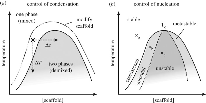
Compartmentalization is a characterizing feature of complexity in cells, used to organize their biochemistry. Membrane-bound organelles are most widely known, but non-membrane-bound liquid organelles also exist. These have recently been shown to form by phase separation of specific types of proteins known as scaffolds. This forms two phases: a condensate that is enriched in scaffold protein separated by a phase boundary from the cytoplasm or nucleoplasm with a low concentration of the scaffold protein. Phase separation is well known for synthetic polymers, but also appears important in cells. Here, we review the properties of proteins important for forming these non-membrane-bound organelles, focusing on the energetically favourable interactions that drive condensation. On this basis we make qualitative predictions about how cells may control compartmentalization by condensates; the partition of specific molecules to a condensate; the control of condensation and dissolution of condensates; and the regulation of condensate nucleation. There are emerging data supporting many of these predictions, although future results may prove incorrect. It appears that many molecules may have the ability to modulate condensate formation, making condensates a potential target for future therapeutics. The emerging properties of condensates are fundamentally unlike the properties of membrane-bound organelles. They have the capacity to rapidly integrate cellular events and act as a new class of sensors for internal and external environments.This article is part of the theme issue 'Self-organization in cell biology'.

Keywords: biomolecular condensates; cell compartmentalization; liquid–liquid phase separation; non-membrane-bound organelle.


Conflict of interest statement

We declare we have no competing interests.

Figures

Figure 1.
Figure 1.
Equilibria underlying control of condensation, solvation and miscibility. Simplified models illustrating mechanisms controlling condensate dynamics and composition. (a) A scaffold protein is present at a low concentration in solution in dynamic equilibrium with the condensate. Conversion to a modified scaffold (increased k1) that cannot form a condensate will change the equilibrium, causing condensate dissolution. The reverse (increased k2) will cause condensation. (b) Control of partition of a molecule into a condensate can be viewed as control of solvation of the molecule by the scaffold in the condensate. A solute is present in dynamic equilibrium between the surrounding solution and the condensate, at a concentration ratio dependent on the partition coefficient. Conversion of the solute to a modified form that is insoluble in the condensate (increased k1) will cause partition away from the condensate. The reverse (increased k2) causes partition to the condensate. (c) One way in which multiple classes of condensates may be controlled through miscibility/immiscibility. A condensate made up of two miscible scaffolds exists in dynamic equilibrium with the low concentration of each scaffold in the surrounding solution. Conversion of scaffold 2 to a modified scaffold 2 (increased k1) will cause partition of scaffold 2 from the droplet (as in a). The modified scaffold 2 may be able to form a second condensate immiscible with the first, and increased k1 would promote this. The reverse would occur on increased k2.
Figure 2.
Figure 2.
Methods to control condensation of a condensate. (a) Three example mechanisms for causing condensation for a cell with starting temperature and scaffold concentration indicated with a cross. The temperature shift ΔT would change the conditions to cross the phase boundary to a region allowing phase separation. Similarly, scaffold concentration change Δc would allow phase separation. Alternatively, modification of the scaffold to change the phase diagram and move the phase boundary such that the current conditions cause demixing would also allow condensate condensation. (b) Under some conditions a demixed state is stable, but condensates do not spontaneously form. At point a the one phase mixed state is stable. At point b, across the phase boundary/coexistence curve, the one phase mixed state is metastable. Given a nucleator, demixing will occur. At point c, across the spinodal curve, the one phase mixed state is unstable and condensates will spontaneously nucleate.

References

    1. Brangwynne CP, Mitchison TJ, Hyman AA. 2011. Active liquid-like behavior of nucleoli determines their size and shape in Xenopus laevis oocytes. Proc. Natl Acad. Sci. USA 108, 4334–4339. (10.1073/pnas.1017150108) - DOI - PMC - PubMed
    1. Brangwynne CP, Eckmann CR, Courson DS, Rybarska A, Hoege C, Gharakhani J, Jülicher F, Hyman AA. 2009. Germline P granules are liquid droplets that localize by controlled dissolution/condensation. Science 324, 1729–1732. (10.1126/science.1172046) - DOI - PubMed
    1. Patel A, et al. 2015. A liquid-to-solid phase transition of the ALS protein FUS accelerated by disease mutation. Cell 162, 1066–1077. (10.1016/j.cell.2015.07.047) - DOI - PubMed
    1. Li P, et al. 2012. Phase transitions in the assembly of multivalent signalling proteins. Nature 483, 336–340. (10.1038/nature10879) - DOI - PMC - PubMed
    1. Su X, et al. 2016. Phase separation of signaling molecules promotes T cell receptor signal transduction. Science 352, 595–599. (10.1126/science.aad9964) - DOI - PMC - PubMed

Publication types