. 2018 Oct;32(10):2250-2262.
doi: 10.1038/s41375-018-0104-2. Epub 2018 Mar 22.

Targeting the MALAT1/PARP1/LIG3 complex induces DNA damage and apoptosis in multiple myeloma

Affiliations

Targeting the MALAT1/PARP1/LIG3 complex induces DNA damage and apoptosis in multiple myeloma

Yi Hu et al. Leukemia. 2018 Oct.

Abstract

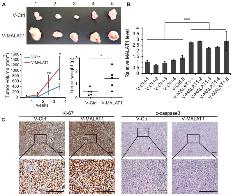
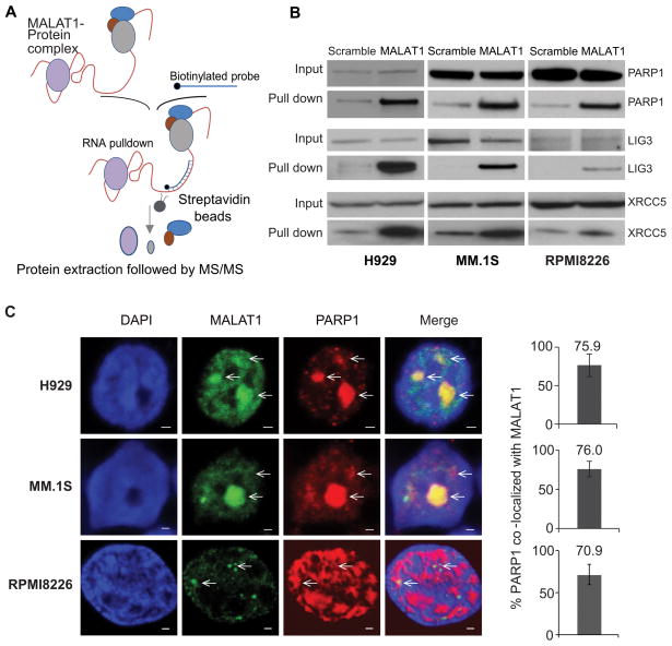
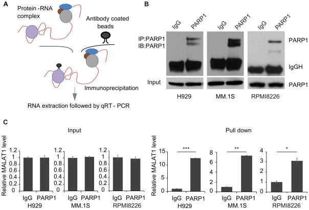
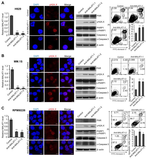
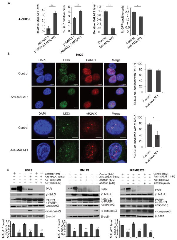
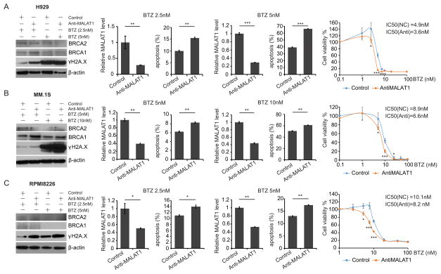
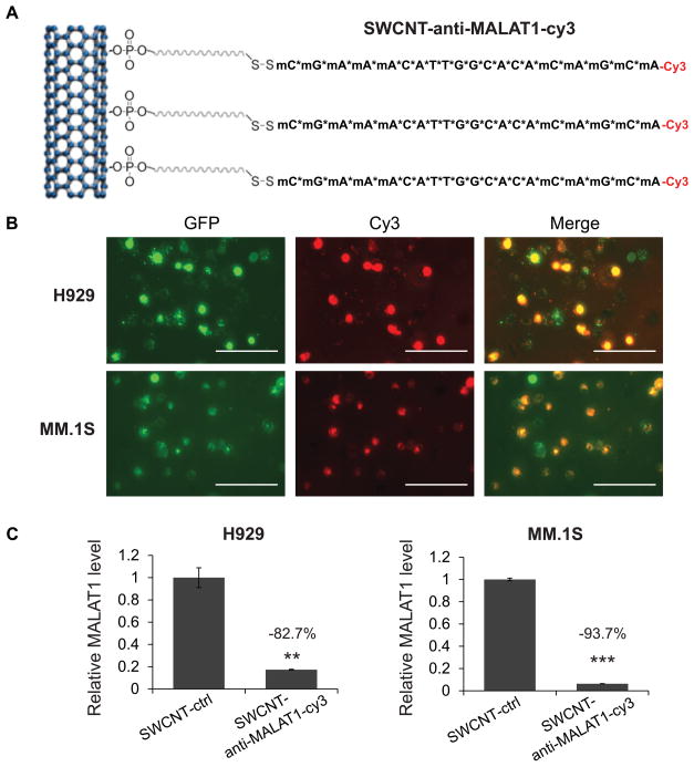
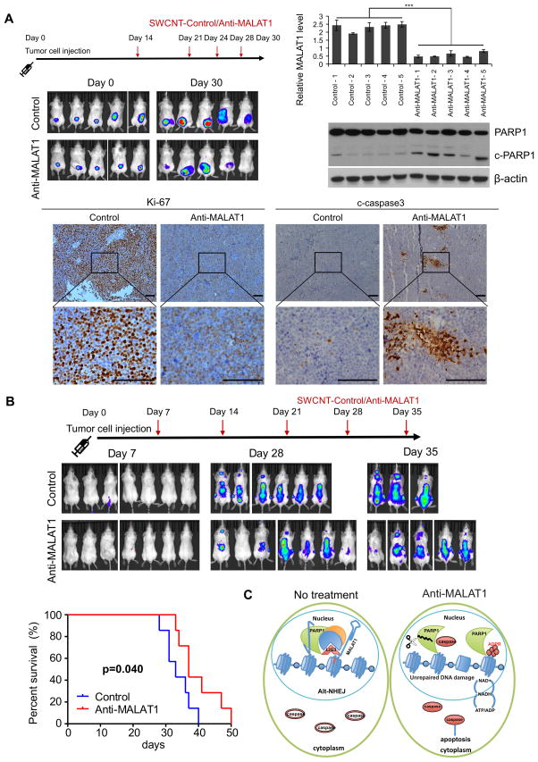
Metastasis-associated lung adenocarcinoma transcript 1 (MALAT1) is a highly conserved long non-coding RNA (lncRNA). Overexpression of MALAT1 has been demonstrated to related to poor prognosis of multiple myeloma (MM) patients. Here, we demonstrated that MALAT1 plays important roles in MM DNA repair and cell death. We found bone marrow plasma cells from patients with monoclonal gammopathy of undetermined significance (MGUS) and MM express elevated MALAT1 and involve in alternative non-homozygous end joining (A-NHEJ) pathway by binding to PARP1 and LIG3, two key components of the A-NHEJ protein complex. Degradation of the MALAT1 RNA by RNase H using antisense gapmer DNA oligos in MM cells stimulated poly-ADP-ribosylation of nuclear proteins, defected the DNA repair pathway, and further provoked apoptotic pathways. Anti-MALAT1 therapy combined with PARP1 inhibitor or proteasome inhibitor in MM cells showed a synergistic effect in vitro. Furthermore, using novel single-wall carbon nanotube (SWCNT) conjugated with anti-MALAT1 oligos, we successfully knocked-down MALAT1 RNA in cultured MM cell lines and xenograft murine models. Most importantly, anti-MALAT1 therapy induced DNA damage and cell apoptosis in vivo, indicating that MALAT1 could serve as a potential novel therapeutic target for MM treatment.


Conflict of interest statement

Conflict-of-interest disclosure: The authors report no potential conflicts of interest.

Figures

Fig. 1
Fig. 1. MALAT1 overexpression promoted the tumorigenesis of MM
(A) 2×10⁶ MALAT1 overexpressed or control MM.1S cells were injected subcutaneously to the shoulder of SCID mice. The sizes of xenograft were measured once a week. Mice were sacrificed 30 days after injection, and xenografts were weighted. (B) MALAT1 level was determined by qRT-PCR. (C) The levels of Ki-67 and c-Caspase3 were detected by immunohistochemistry. (*p<0.05, **p<0.01, ***p<0.001)
Fig. 2
Fig. 2. PARP1/LIG3 complex was identified as MALAT1 binding target by RAP-MS
(A) Schematic diagram of the RAP-MS strategy used to identify MALAT1 binding proteins. (B) PARP1, LIG3 and XRCC5 were verified as MALAT1 binding proteins. (C) MALAT1 co-localized with PARP1 in H929, MM.1S and RPMI8226 cells (scale bar=1μM).
Fig. 3
Fig. 3. Verification of the binding between MALAT1 and PARP1 by RIP-PCR
(A) Schematic diagram of the RIP-PCR assay. (B) The level of PARP1 before or after RIP was determined by immunoblotting. (C) MALAT1 pulled-down by PARP1 antibody-conjugated beads were determined by qRT-PCR (right panel), left panel is input (*p<0.05, **p<0.01, ***p<0.001).
Fig. 4
Fig. 4. MALAT1 inhibition induced DNA damage and cell death in MM
2′-OMe-modified anti-MALAT1 oligos or control oligos were transfected into H929(A), MM.1S(B) or RPMI8226(C) cells, respectively. At 48h after transfection, cells were collected and subjected to qRT-PCR, immunofluorescence staining for γH2A.X, immunoblotting of γH2A.X, PARP1, c-PARP1, caspase-3, and c-caspase3 and apoptosis assay. (*p<0.05, **p<0.01).
Fig. 5
Fig. 5. MALAT1 coordinated with PAPR1 inhibitor through inhibiting A-NHEJ
(A) In A-NHEJ reporter plasmid (pEJ2-GFP-puro) stable expression 293T cells, pCBA-SceI was transient transfected with MALAT1 overexpression/empty vector or anti-MALAT1/control gapmer. Then GFP positive cells were determined by flow cytometry. (B) The co-localization between LIG3 and PARP1 or γH2A.X were determined by immunofluorescence staining (scale bar=5μM). (C) H929, MM.1S and RPMI8226 cells transfected with anti-MALAT1 or control oligos were treated with ABT-888. Cells were collected for WB. MALAT1 were determined by qRT-PCR.(*p<0.05, **p<0.01)
Fig. 6
Fig. 6. Synergistic effect of anti-MALAT1 and bortezomib in MM
H929(A), MM.1S(B) and RPMI8226(C) cells were transfected with 1nM anti-MALAT1 or control oligos and treated with bortezomib. Cells were collected for immunoblotting, apoptosis assay and qRT-PCR. Cell viability was measured and IC50 was calculated before and after MALAT1 knockdown.( *p<0.05, **p<0.01, ***p<0.001)
Fig. 7
Fig. 7. SWCNT-anti-MALAT1 showed high delivery efficiency and minimal toxicity
(A) Schematic diagram of SWCNT-anti-MALAT1-Cy3 gapmer oligos (Scale bars=100μM). (B) H929-GFP and MM.1S-GFP cells were co-cultured with SWCNT-anti-MALAT1-cy3 for 48h. (C) MALAT1 level was knocked-down successfully.(**p<0.01, ***p<0.001).
Fig. 8
Fig. 8. SWCNT-anti-MALAT1 treatment repressed myeloma growth in both xenograft and disseminated murine models
(A) MM.1S-Luc-GFP cells were injected subcutaneously to SCID mice (5 mice each group). SWCNT-anti-MALAT1 or SWCNT-ctrl was injected into the tumors at the indicated days. Tumor growth was monitored by IVIS. Mice were sacrificed on day 30, tumor samples were subjected to qRT-PCR, WB and immunohistochemistry (Scale bars=100μM). (B) SCID mice (7 mice each group) were intravenously injected with MM.1S-Luc-GFP cells, then injected with SWCNT-anti-MALAT1 or SWCNT-ctrl once a week through tail veins. Kaplan-Meier analysis indicated SWCNT-anti-MALAT1 prolonged mouse lifespan significantly (P=0.04). (C) Proposed model of MALAT1 antagonist induces MM cell apoptosis.

References

    1. Siegel R, Naishadham D, Jemal A. Cancer statistics, 2013. CA: a cancer journal for clinicians. 2013 Jan;63(1):11–30. - PubMed
    1. Ntziachristos P, Abdel-Wahab O, Aifantis I. Emerging concepts of epigenetic dysregulation in hematological malignancies. Nature immunology. 2016 Sep;17(9):1016–1024. - PMC - PubMed
    1. Evans JR, Feng FY, Chinnaiyan AM. The bright side of dark matter: lncRNAs in cancer. The Journal of clinical investigation. 2016 Aug 01;126(8):2775–2782. - PMC - PubMed
    1. Ronchetti D, Agnelli L, Taiana E, Galletti S, Manzoni M, Todoerti K, et al. Distinct lncRNA transcriptional fingerprints characterize progressive stages of multiple myeloma. Oncotarget. 2016 Mar 22;7(12):14814–14830. - PMC - PubMed
    1. Wong KY, Li Z, Zhang X, Leung GK, Chan GC, Chim CS. Epigenetic silencing of a long non-coding RNA KIAA0495 in multiple myeloma. Molecular cancer. 2015 Sep 26;14:175. - PMC - PubMed

Publication types