. 2018 Mar 26:12:82.
doi: 10.3389/fncel.2018.00082. eCollection 2018.

The Enlightened Brain: Novel Imaging Methods Focus on Epileptic Networks at Multiple Scales

Affiliations

The Enlightened Brain: Novel Imaging Methods Focus on Epileptic Networks at Multiple Scales

L Federico Rossi et al. Front Cell Neurosci. .

Abstract

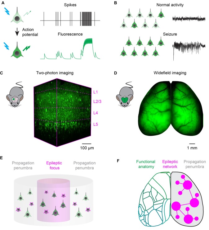
Epilepsy research is rapidly adopting novel fluorescence optical imaging methods to tackle unresolved questions on the cellular and circuit mechanisms of seizure generation and evolution. State of the art two-photon microscopy and wide-field fluorescence imaging can record the activity in epileptic networks at multiple scales, from neuronal microcircuits to brain-wide networks. These approaches exploit transgenic and viral technologies to target genetically encoded calcium and voltage sensitive indicators to subclasses of neurons, and achieve genetic specificity, spatial resolution and scalability that can complement electrophysiological recordings from awake animal models of epilepsy. Two-photon microscopy is well suited to study single neuron dynamics during interictal and ictal events, and highlight the differences between the activity of excitatory and inhibitory neuronal classes in the focus and propagation zone. In contrast, wide-field fluorescence imaging provides mesoscopic recordings from the entire cortical surface, necessary to investigate seizure propagation pathways, and how the unfolding of epileptic events depends on the topology of brain-wide functional connectivity. Answering these questions will inform pre-clinical studies attempting to suppress seizures with gene therapy, optogenetic or chemogenetic strategies. Dissecting which network nodes outside the seizure onset zone are important for seizure generation, propagation and termination can be used to optimize current and future evaluation methods to identify an optimal surgical strategy.

Keywords: 2-photon imaging; epilepsy; in vivo imaging; seizure; wide-field Ca2+ imaging.


Figures

Figure 1
Figure 1
Fluorescence imaging of epileptic activity at multiple scales. (A) Fluorescent activity reporters can be targeted to neurons to read out their activity. Reporters change the intensity, or wavelength, of their fluorescence as a function of neuronal activity. (B) Fluorescent reporters targeted to neurons at the population level read out the hallmarks of epileptic activity. During normal processing (top), neuronal activity is sparse and EEG is desynchronized. During seizures, activity escalates recruiting most of the neurons in the network and generates hypersynchronous EEG oscillations. (C) Two-photon imaging can be used to record neuronal activity with single cell resolution in a small cortical volume. 3D-reconstruction of a 400 × 800 × 650 μm volume recorded in vivo in the visual cortex of a transgenic mouse expressing the genetically encoded calcium indicator GCaMP6s in neocortical excitatory neurons across layers (magenta dotted lines). (D) Wide-field imaging can be used to record neuronal activity over the entire dorsal cortex of rodents. Example fluorescence image of the dorsal cortex from a mouse expressing the genetically encoded calcium indicator GCaMP6s in all neocortical excitatory neurons (courtesy of Dr. Nicholas A. Steinmetz). (E) Two-photon microscopy can record single neuron dynamics during interictal and ictal discharges, and highlight the differences between the activity of excitatory (green) and inhibitory (magenta) neuronal classes in the focus and propagation penumbra. (F) Wide-field fluorescence imaging provides mesoscopic mapping of the functional anatomy of the cortex (green outlines, current neuro-anatomical subdivisions of mouse cortex from the Allen Brain Atlas); it can investigate seizure propagation pathways and ask how the unfolding of epileptic events depends on the topology of brain wide functional connectivity.

References

    1. Aiba I., Noebels J. L. (2015). Spreading depolarization in the brainstem mediates sudden cardiorespiratory arrest in mouse SUDEP models. Sci. Transl. Med. 7:282ra46. 10.1126/scitranslmed.aaa4050 - DOI - PMC - PubMed
    1. Andermann M. L., Gilfoy N. B., Goldey G. J., Sachdev R. N. S., Wölfel M., McCormick D. A., et al. . (2013). Chronic cellular imaging of entire cortical columns in awake mice using microprisms. Neuron 80, 900–913. 10.1016/j.neuron.2013.07.052 - DOI - PMC - PubMed
    1. Arosio D., Ratto G. M. (2014). Twenty years of fluorescence imaging of intracellular chloride. Front. Cell. Neurosci. 8:258. 10.3389/fncel.2014.00258 - DOI - PMC - PubMed
    1. Baird-Daniel E., Daniel A. G. S., Wenzel M., Li D., Liou J., Laffont P., et al. . (2017). Glial calcium waves are triggered by seizure activity and not essential for initiating ictal onset or neurovascular coupling. Cereb. Cortex 27, 3318–3330. 10.1093/cercor/bhx072 - DOI - PMC - PubMed
    1. Barretto R. P. J., Messerschmidt B., Schnitzer M. J. (2009). In vivo fluorescence imaging with high-resolution microlenses. Nat. Methods 6, 511–512. 10.1038/nmeth.1339 - DOI - PMC - PubMed

LinkOut - more resources