

Molecular Characterization of a Dual Domain Carbonic Anhydrase From the Ctenidium of the Giant Clam, Tridacna squamosa, and Its Expression Levels After Light Exposure, Cellular Localization, and Possible Role in the Uptake of Exogenous Inorganic Carbon
- PMID: 29632495
- PMCID: PMC5879104
- DOI: 10.3389/fphys.2018.00281
Molecular Characterization of a Dual Domain Carbonic Anhydrase From the Ctenidium of the Giant Clam, Tridacna squamosa, and Its Expression Levels After Light Exposure, Cellular Localization, and Possible Role in the Uptake of Exogenous Inorganic Carbon
Abstract
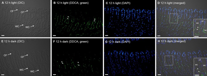
A Dual-Domain Carbonic Anhydrase (DDCA) had been sequenced and characterized from the ctenidia (gills) of the giant clam, Tridacna squamosa, which lives in symbiosis with zooxanthellae. DDCA was expressed predominantly in the ctenidium. The complete cDNA coding sequence of DDCA from T. squamosa comprised 1,803 bp, encoding a protein of 601 amino acids and 66.7 kDa. The deduced DDCA sequence contained two distinct α-CA domains, each with a specific catalytic site. It had a high sequence similarity with tgCA from Tridacna gigas. In T. squamosa, the DDCA was localized apically in certain epithelial cells near the base of the ctenidial filament and the epithelial cells surrounding the tertiary water channels. Due to the presence of two transmembrane regions in the DDCA, one of the Zn2+-containing active sites could be located externally and the other one inside the cell. These results denote that the ctenidial DDCA was positioned to dehydrate [Formula: see text] to CO2 in seawater, and to hydrate the CO2 that had permeated the apical membrane back to [Formula: see text] in the cytoplasm. During insolation, the host clam needs to increase the uptake of inorganic carbon from the ambient seawater to benefit the symbiotic zooxanthellae; only then, can the symbionts conduct photosynthesis and share the photosynthates with the host. Indeed, the transcript and protein levels of DDCA/DDCA in the ctenidium of T. squamosa increased significantly after 6 and 12 h of exposure to light, respectively, denoting that DDCA could participate in the light-enhanced uptake and assimilation of exogenous inorganic carbon.
Keywords: Symbiodinium; bicarbonate; calcification; carbon dioxide; symbiosis; tridacnid.
Figures


Similar articles
-
Calcium absorption in the fluted giant clam, Tridacna squamosa, may involve a homolog of voltage-gated calcium channel subunit α1 (CACNA1) that has an apical localization and displays light-enhanced protein expression in the ctenidium.J Comp Physiol B. 2019 Dec;189(6):693-706. doi: 10.1007/s00360-019-01238-4. Epub 2019 Oct 4. J Comp Physiol B. 2019. PMID: 31586259
-
Molecular characterization, light-dependent expression, and cellular localization of a host vacuolar-type H+-ATPase (VHA) subunit A in the giant clam, Tridacna squamosa, indicate the involvement of the host VHA in the uptake of inorganic carbon and its supply to the symbiotic zooxanthellae.Gene. 2018 Jun 15;659:137-148. doi: 10.1016/j.gene.2018.03.054. Epub 2018 Mar 17. Gene. 2018. PMID: 29559349
-
Carbonic anhydrase 2-like in the giant clam, Tridacna squamosa: characterization, localization, response to light, and possible role in the transport of inorganic carbon from the host to its symbionts.Physiol Rep. 2017 Dec;5(23):e13494. doi: 10.14814/phy2.13494. Physiol Rep. 2017. PMID: 29199178 Free PMC article.
-
Light exposure enhances urea absorption in the fluted giant clam, Tridacna squamosa, and up-regulates the protein abundance of a light-dependent urea active transporter, DUR3-like, in its ctenidium.J Exp Biol. 2018 Apr 19;221(Pt 8):jeb176313. doi: 10.1242/jeb.176313. J Exp Biol. 2018. PMID: 29540461
-
Prokaryotic carbonic anhydrases.FEMS Microbiol Rev. 2000 Oct;24(4):335-66. doi: 10.1111/j.1574-6976.2000.tb00546.x. FEMS Microbiol Rev. 2000. PMID: 10978542 Review.
Cited by
-
The ctenidium of the giant clam, Tridacna squamosa, expresses an ammonium transporter 1 that displays light-suppressed gene and protein expression and may be involved in ammonia excretion.J Comp Physiol B. 2018 Sep;188(5):765-777. doi: 10.1007/s00360-018-1161-6. Epub 2018 Apr 24. J Comp Physiol B. 2018. PMID: 29691634
-
Intracellular pH regulation in mantle epithelial cells of the Pacific oyster, Crassostrea gigas.J Comp Physiol B. 2020 Nov;190(6):691-700. doi: 10.1007/s00360-020-01303-3. Epub 2020 Aug 20. J Comp Physiol B. 2020. PMID: 32816118 Free PMC article.
-
Calcium absorption in the fluted giant clam, Tridacna squamosa, may involve a homolog of voltage-gated calcium channel subunit α1 (CACNA1) that has an apical localization and displays light-enhanced protein expression in the ctenidium.J Comp Physiol B. 2019 Dec;189(6):693-706. doi: 10.1007/s00360-019-01238-4. Epub 2019 Oct 4. J Comp Physiol B. 2019. PMID: 31586259
-
The colorful mantle of the giant clam Tridacna squamosa expresses a homolog of electrogenic sodium: Bicarbonate cotransporter 2 that mediates the supply of inorganic carbon to photosynthesizing symbionts.PLoS One. 2021 Oct 15;16(10):e0258519. doi: 10.1371/journal.pone.0258519. eCollection 2021. PLoS One. 2021. PMID: 34653199 Free PMC article.
References
-
- Aizawa K., Miyachi S. (1986). Carbonic anhydrase and CO2 concentrating mechanisms in microalgae and cyanobacteria. FEMS Microbiol. Rev. 39, 215–233. 10.1111/j.1574-6968.1986.tb01860.x - DOI
-
- Al-Moghrabi S., Goiran C., Allemand D., Speziale N., Jaubert J. (1996). Inorganic carbon uptake for photosynthesis by symbiotic coral/dinoflagellate associations. II. Mechanisms for bicarbonate uptake. J. Exp. Mar. Biol. Ecol. 199, 227–248. 10.1016/0022-0981(95)00202-2 - DOI
-
- Badger M. R., Price G. D. (1994). The role of carbonic anhydrase in photosynthesis. Ann. Rev. Plant Physiol. Plant Mol. Biol. 45, 369–392. 10.1146/annurev.pp.45.060194.002101 - DOI
LinkOut - more resources
Full Text Sources
Other Literature Sources