

Lateral acetabular coverage as a predictor of femoroacetabular cartilage thickness
- PMID: 29632686
- PMCID: PMC5883176
- DOI: 10.1093/jhps/hnw034
Lateral acetabular coverage as a predictor of femoroacetabular cartilage thickness
Abstract
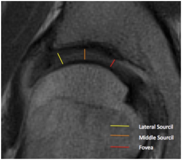
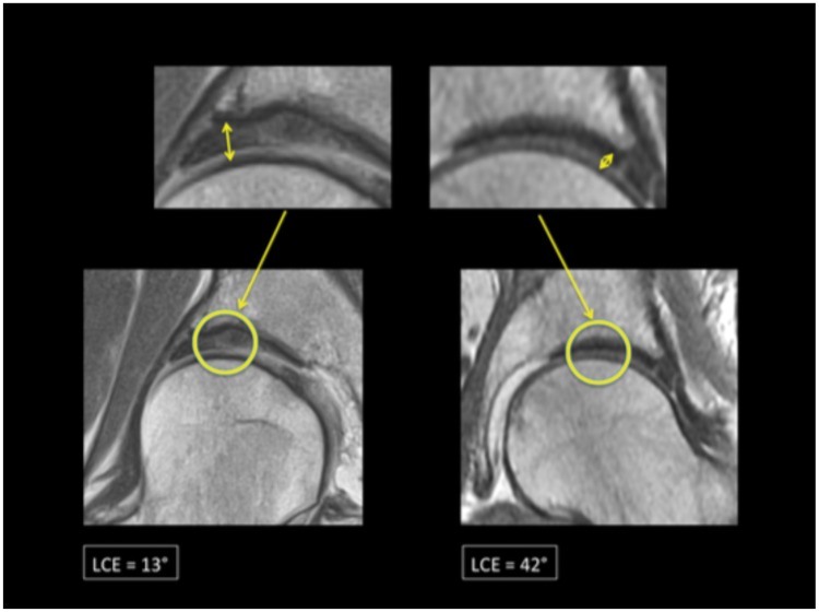
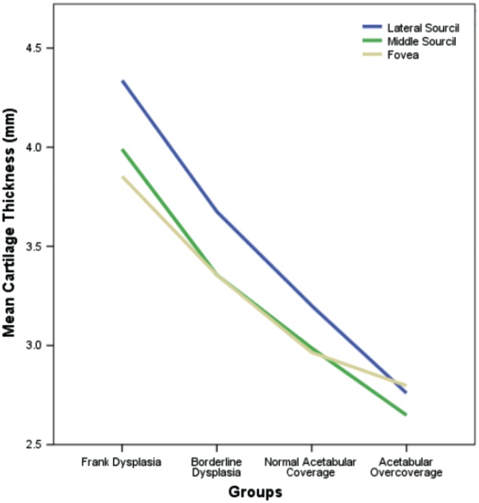
To investigate the correlation between femoroacetabular cartilage thickness and lateral acetabular coverage in patients undergoing hip arthroscopy for a variety of indications. Articular cartilage at the hip is hypothesized to undergo adaptive change secondary to unique patterns of pathomechanical loading which results in a direct relationship between acetabular coverage and femoroacetabular cartilage thickness. A cohort of 252 patients presenting to our dedicated hip preservation service between June 2013 and June 2015 were retrospectively analysed. Preoperative radiographs and MRI studies were obtained for all symptomatic hips and classified according to radiographic lateral center edge angle (LCEA) as follows: normal acetabular coverage (25-40°), acetabular overcoverage (≥40°), borderline dysplasia (20-24.9°) and frank dysplasia (<20°). Femoroacetabular cartilage thickness was measured on a preoperative MRI-scan at the fovea, middle sourcil, and lateral sourcil. In all groups, cartilage thickness was maximized at the lateral sourcil relative to the middle sourcil or fovea (P < 0.001). Furthermore, articular cartilage thickness was significantly increased when comparing one group to successive groups with diminished lateral acetabular coverage. Indeed, multivariate analyses confirmed LCEA to be the strongest determinant of femoroacetabular cartilage thickness compared with age, gender, body-mass index or presence of cam/pincer lesions. Patients with borderline and frank dysplasia exhibit increased values of femoroacetabular cartilage thickness in the weight-bearing zone, potentially indicating a compensatory reaction to the lack of bony coverage. Articular cartilage thickness may serve as an instability marker and inform clinical decision-making for patients with borderline dysplasia.
Figures
References
-
- Crawford MJ, Dy CJ, Alexander JW. et al. The Biomechanics of the Hip Labrum and the Stability of the Hip. Clin Orthop Relat Res 2007; 465:16–22. - PubMed
LinkOut - more resources
Full Text Sources
Other Literature Sources
