Review
. 2018 Sep;96(9):1543-1559.
doi: 10.1002/jnr.24240. Epub 2018 Apr 6.

Embedded ensemble encoding hypothesis: The role of the "Prepared" cell

Affiliations
Review

Embedded ensemble encoding hypothesis: The role of the "Prepared" cell

Srdjan D Antic et al. J Neurosci Res. 2018 Sep.

Abstract

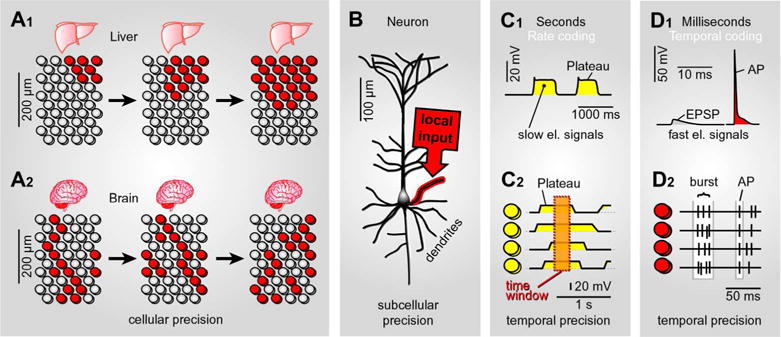
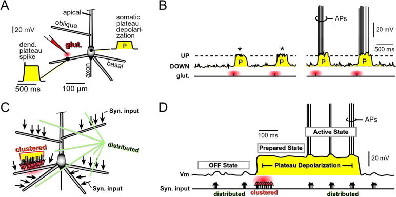
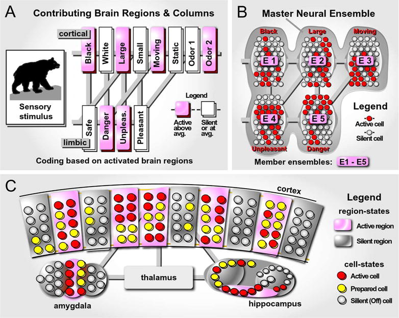
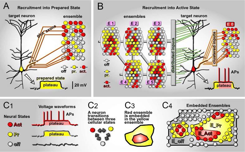
We here reconsider current theories of neural ensembles in the context of recent discoveries about neuronal dendritic physiology. The key physiological observation is that the dendritic plateau potential produces sustained depolarization of the cell body (amplitude 10-20 mV, duration 200-500 ms). Our central hypothesis is that synaptically-evoked dendritic plateau potentials lead to a prepared state of a neuron that favors spike generation. The plateau both depolarizes the cell toward spike threshold, and provides faster response to inputs through a shortened membrane time constant. As a result, the speed of synaptic-to-action potential (AP) transfer is faster during the plateau phase. Our hypothesis relates the changes from "resting" to "depolarized" neuronal state to changes in ensemble dynamics and in network information flow. The plateau provides the Prepared state (sustained depolarization of the cell body) with a time window of 200-500 ms. During this time, a neuron can tune into ongoing network activity and synchronize spiking with other neurons to provide a coordinated Active state (robust firing of somatic APs), which would permit "binding" of signals through coordination of neural activity across a population. The transient Active ensemble of neurons is embedded in the longer-lasting Prepared ensemble of neurons. We hypothesize that "embedded ensemble encoding" may be an important organizing principle in networks of neurons.

Keywords: UP state; binding; dendritic; glutamate; plateau potential; rate code.


Conflict of interest statement

Conflict of Statement

The authors declare no conflicts of interest.

Figures

Fig. 1
Fig. 1. Unique advantages of electrical signaling
(A1) Schematic representation of a liver tissue. Each circle represents one hepatocyte. Color indicates “activated” cells. Cellular activity is synchronized among neighboring hepatocytes by diffusing chemical signals. (A2) Schematic representation of a brain tissue section. Each circle represents one neuron. Color indicates “activated” cells. Cellular activity is synchronized between particular neurons (not all neurons) by electrical signaling. (B) Synaptic inputs impinge on one basal dendrite of a cortical pyramidal cell. Synaptically-induced depolarization is strongest in one cellular compartment (red halo). (C1) Slow electrical signals in the form of sustained plateau depolarizations; amplitude ~20 mV, duration ~500 ms. (C2) Electrical activity is synchronized among 4 cells on a slow temporal scale (seconds). Dashed rectangle indicates a period of time when all 4 cells are in depolarized state. (D1) Fast electrical signals, excitatory postsynaptic potential (EPSP) and action potential (AP), are shown on a fast time scale. (D2) Action potential firing is simultaneously recorded in 4 neurons. Each vertical tick represents one AP, as in “single-unit” recordings. Synchronization can be based on the temporal relation between between groups of APs (AP bursts) or individual APs (spikes). Synchronized bursts of APs or synchronized single APs are marked by white background rectangle.
Fig. 2
Fig. 2. Dendritic plateau potentials – somatic plateau depolarizations
(A) Cartoon of a pyramidal neuron showing classes of dendrites: basal, oblique and apical trunk. Voltage waveform of dendritic plateau potential and the resulting somatic depolarization (“P”), in response to glutamatergic stimulation of one basal dendrite. “Glut.” marks the glutamate iontophoresis site. (B) Cartoon of four glutamate iontophoresis pulses (each pulse = 5 ms) delivered on one basal dendrite. Glutamate stimuli trigger somatic plateau depolarizations (P), which resemble neuronal UP states. The somatic P without APs (*) is termed a “Prepared state” of a neuron (Pr). The somatic P accompanied by action potentials (APs) is termed “Active state” (Act). The somatic DOWN state is termed an “OFF state” of a neuron (off). (C) Cartoon of a pyramidal neuron - black arrows mark glutamatergic inputs of approximately identical weight. Two spatial patterns of synaptic inputs are shown: distributed (everywhere) and clustered (on one basal dendrite). Clustered inputs produce glutamate spillover (red cloud). (D) At the low levels of incoming synaptic inputs, the somatic membrane potential (Vm) is dwelling near resting potential (OFF state). Clustered glutamatergic inputs onto one basal dendrite (“clustered”) produce spillover glutamate (red cloud), which triggers dendritic plateau potential, which in turn brings the cell body into a plateau depolarization. During the plateau depolarization (Prepared state) the membrane potential is ~20 mV closer to the AP threshold and the membrane time constant is shorter due to a glutamate-mediated drop in membrane resistance. As a result, the neuron is more responsive to incoming synaptic inputs distributed across the dendritic tree (black arrows). The same-size synaptic input (3 arrows) fails to initiate AP in the OFF state, but successfully drives AP firing in the Prepared state.
Fig. 3
Fig. 3. Object coding by neural ensembles
(A) An object in the perceptual field (bear) activates pockets of cells in many brain regions. Each rectangle represents a column, or a segment of a nucleus, devoted to one simple attribute of the object. Brain regions are connected with axonal projections (gray stripes). The overall activity of a column increases above average if an attribute were detected (pink) – the column is “net-active”. If an attribute was not detected, the net electrical activity remains at or below the average level (white) – the column is “net-silent”. (B) Schematic depiction of net active brain regions at cellular resolution. The net silent brain regions (white rectangles in the previous panel) are omitted form this presentation, for simplicity. Active neurons = red circles. Inactive neurons = gray circles. Multiple neural ensembles (E1 – E5) are embedded in the master neural ensemble, which codes for a large, dark and dangerous moving object detected by sensory system. Member ensembles (E1 – E5) may reside in separate brain regions (e.g. cortex, striatum and amygdala). Together the member ensembles form a master neural ensemble (gray contour). (C) Schematic depiction of 4 brain areas: cortex, hippocampus, thalamus and amygdala. Each brain area is divided into smaller functional units/segments/columns. Units can be either net-active (pink) or net-silent (gray). In respect to electrical activity at single-cell level, neurons can be found in 3 functional states: OFF (gray circle), Prepared (yellow circle) and Active (red circle).
Fig. 4
Fig. 4. Recruitment of neurons into neural ensembles
(A) A pyramidal neuron is receiving glutamatergic projections from a brain region comprised of neurons in three characteristic states of activity (OFF, Pr and Act). A fraction of spiking cells (red) sends their glutamatergic projections (glut. axons) onto the target pyramidal neuron, clustering on one particular basal branch. A computational task engaging the neural ensemble (red cell ensemble activity) is poised to recruit the target neuron into a Prepared state (somatic plateau depolarization, amplitude ≈ 20 mV, duration ≈ 300 ms), via the generation of a local dendritic plateau potential (basal dendrite - orange halo). (B) A target pyramidal cell is receiving two types of glutamatergic inputs, clustered and distributed. “Clustered inputs” arrive from ensemble E0. Thus, ensemble E0 has the capacity to recruit our neuron into Prepared state, by triggering local dendritic plateau potential (same as in A). “Distributed inputs” arrive from various active brain regions (E1 – E5) and they scatter across the entire dendritic tree, including basal, oblique and apical tuft branches. Distributed inputs have the capacity to drive AP firing, but only if E0 had been successful in recruiting this cell into a Prepared state. (C1) A characteristic somatic voltage waveform applies to each neuronal state, OFF, Prepared and Active. (C2) Each neuron transitions between 3 basic states, depending on the ongoing pattern of excitatory glutamatergic inputs. (C3) A group of Active cells (red contour) are recruited from a group of Prepared cells (yellow contour). (C4) Cartoon of a brain region shown at “cellular resolution”, each circle represents one neuron. The ensemble made of neurons in Active state (E_Act, red) is just a subset of the ensemble of neurons in Prepared state (E_Pr, yellow), which itself is a subset of neurons in the OFF state (E_off, gray).

References

    1. Agmon-Snir H, Carr CE, Rinzel J. The role of dendrites in auditory coincidence detection. Nature. 1998;393:268–272. - PubMed
    1. Ahissar E, Sosnik R, Haidarliu S. Transformation from temporal to rate coding in a somatosensory thalamocortical pathway. Nature. 2000;406:302–306. - PubMed
    1. Ainsworth M, Lee S, Cunningham MO, Traub RD, Kopell NJ, Whittington MA. Rates and rhythms: a synergistic view of frequency and temporal coding in neuronal networks. Neuron. 2012;75:572–583. - PubMed
    1. Allen WE, Kauvar IV, Chen MZ, Richman EB, Yang SJ, Chan K, Gradinaru V, Deverman BE, Luo L, Deisseroth K. Global Representations of Goal-Directed Behavior in Distinct Cell Types of Mouse Neocortex. Neuron. 2017;94:891–907 e896. - PMC - PubMed
    1. Antic SD, Empson RM, Knopfel T. Voltage imaging to understand connections and functions of neuronal circuits. J Neurophysiol. 2016;116:135–152. - PMC - PubMed

Publication types