. 2018 Apr 6;109(1):41-62.
doi: 10.1111/mmi.13966. Online ahead of print.

Variant-specific and reciprocal Hsp40 functions in Hsp104-mediated prion elimination

Affiliations

Variant-specific and reciprocal Hsp40 functions in Hsp104-mediated prion elimination

Michael T Astor et al. Mol Microbiol. .

Abstract

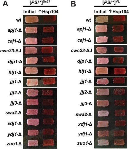
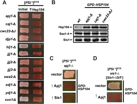
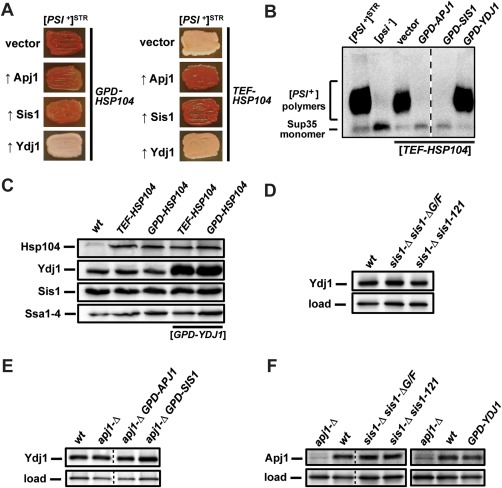
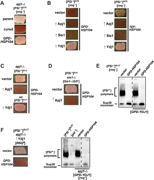
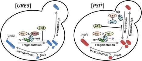
The amyloid-based prions of Saccharomyces cerevisiae are heritable aggregates of misfolded proteins, passed to daughter cells following fragmentation by molecular chaperones including the J-protein Sis1, Hsp70 and Hsp104. Overexpression of Hsp104 efficiently cures cell populations of the prion [PSI+ ] by an alternative Sis1-dependent mechanism that is currently the subject of significant debate. Here, we broadly investigate the role of J-proteins in this process by determining the impact of amyloid polymorphisms (prion variants) on the ability of well-studied Sis1 constructs to compensate for Sis1 and ask whether any other S. cerevisiae cytosolic J-proteins are also required for this process. Our comprehensive screen, examining all 13 members of the yeast cytosolic/nuclear J-protein complement, uncovered significant variant-dependent genetic evidence for a role of Apj1 (antiprion DnaJ) in this process. For strong, but not weak [PSI+ ] variants, depletion of Apj1 inhibits Hsp104-mediated curing. Overexpression of either Apj1 or Sis1 enhances curing, while overexpression of Ydj1 completely blocks it. We also demonstrated that Sis1 was the only J-protein necessary for the propagation of at least two weak [PSI+ ] variants and no J-protein alteration, or even combination of alterations, affected the curing of weak [PSI+ ] variants, suggesting the possibility of biochemically distinct, variant-specific Hsp104-mediated curing mechanisms.


Figures

Figure 1
Figure 1
J‐protein primary structure diagrams and Sis1/Droj1 sequence alignment. A. Comparison of primary structures of J‐proteins and J‐protein constructs used in this study. Protein regions are denoted using the following notation: J, J‐domain; G/F, glycine/phenylalanine‐rich region; G/M, glycine/methionine‐rich region; ZBD, zinc binding domain, CTD I/II, C‐terminal peptide‐binding domains I and II; D, dimerization domain. Lines indicate where a region has been deleted. B. Primary sequence alignment between Sis1 (top) and Droj1 (bottom) using the Jotun Hein algorithm of MegAlign from DNAstar (DNASTAR, Madison, WI). Identical residues are highlighted. Numbers above the sequence refer to residue positions in the consensus sequence; numbers to the right indicate residues in each protein.
Figure 2
Figure 2
Droj1 supports strong, but not weak, [PSI+] variants. A. [PSI +] cells of the W303 (left column) and 74D‐694 (right column) genetic backgrounds lacking genomic Sis1 but expressing Sis1 from a URA3‐marked plasmid were transformed with a plasmid expressing Droj1 (GPD‐DROJ1). Following loss of the URA3‐marked plasmid, cells were passaged onto rich medium to test for prion maintenance. Across both backgrounds, four different [PSI +] variants, two strong and two weak, were examined. Color phenotype assays are shown for representative transformants (n ≥ 12 for each strain). B. Cell lysates of strains bearing strong [PSI+] variants Sc4 and VH in the W303 and 74D‐694 yeast genetic backgrounds expressing either Sis1 or Droj1 were resolved by SDD‐AGE and subjected to immunoblot analysis using an antibody specific to Sup35.
Figure 3
Figure 3
Droj1 is deficient in supporting Hsp104‐mediated [PSI+] curing. A. Strong [PSI +] variants of the W303 (top half) and 74D‐694 (bottom half) genetic backgrounds possessing either a plasmid expressing Sis1 (SIS1‐SIS1; left half) or a plasmid expressing Droj1 (GPD‐DROJ1; right half) in place of Sis1 were passaged onto rich medium (left columns). Cells were then transformed with a plasmid overexpressing Hsp104 (GPD‐HSP104) that normally cures [PSI +] (right columns). Color phenotype assays are shown for representative transformants (n ≥ 12 for each strain). B. Lysates of representative cells from the right side of panel A were resolved by SDD‐AGE and subjected to immunoblot analysis using an antibody specific to Sup35. C. Lysates of representative cells from panel B were resolved by SDS‐PAGE and subjected to immunoblot analysis using an antibody specific to Hsp104. Load control shown is a nonspecific protein cross‐reacting with our Hsp104 primary antibody.
Figure 4
Figure 4
Sis1 domain requirements for Hsp104‐mediated curing of strong and weak [PSI +] variants. A. Strong [PSI +]Sc4 cells of the W303 genetic background lacking genomic Sis1 but expressing Sis1 from a URA3‐marked plasmid were transformed with plasmids expressing wild‐type Sis1 or Sis1 truncations Sis1‐ΔG/F or Sis1–121 (left column). Following loss of the URA3‐marked plasmid, cells were then transformed with a plasmid overexpressing Hsp104 (GPD‐HSP104) that normally cures [PSI +]. Color phenotype assays are shown for representative transformants (n ≥ 10 for each variant). B. Lysates of strains lacking wild‐type Sis1 expression (sis1‐Δ) and expressing Sis1, Sis1‐ΔG/F or Sis1–121 from plasmids were resolved by SDS‐PAGE and subjected to immunoblot analysis using an antibody specific for Hsp104. Antibody specific for Ssc1 was used as a loading control. C. Same as panel A, but cells have the weak variant [PSI +]Sc37, and Sis1–121 is omitted because it is unable to propagate this variant. D. Same as panels A and C, but cells are derived from the 74D‐694 genetic background.
Figure 5
Figure 5
No cytosolic J‐protein other than Sis1 is required for propagation or Hsp104‐mediated curing of two weak [PSI +] variants. Cells of the W303 genetic background were used which harbor either the weak [PSI +] variant [PSI +]Sc37 (A), or the weak variant [PSI +]VL (B). Weak [PSI +] bearing cells lacking individual J‐proteins were passaged onto rich medium (left columns). Cells were then transformed with a plasmid overexpressing Hsp104 (GPD‐HSP104) that normally cures [PSI +] (right columns). Color phenotype assays are shown for representative transformants (n ≥ 10 for each variant).
Figure 6
Figure 6
Lack of Apj1 expression, but not any of 11 other cytosolic J‐proteins, impairs Hsp104 curing of strong [PSI +]STR. A. Strong [PSI +]STR bearing cells of the W303 genetic background lacking individual J‐proteins were passaged onto rich medium (left columns). Cells were then transformed with a plasmid overexpressing Hsp104 (GPD‐HSP104) that normally cures [PSI +] (right columns). Color phenotype assays are shown for representative transformants: for apj1‐Δ 48 out of 90 transformants remained [PSI +]; for all other strains curing was complete with n ≥ 10. B. Lysates of a wild‐type strain (wt), a strain lacking Apj1 expression (apj1‐Δ), a strain lacking Caj1 expression (caj1‐Δ) and a strain lacking the J‐domain of Cwc23 (cwc23‐ΔJ) were resolved by SDS‐PAGE and subjected to immunoblot analysis using antibodies specific for Hsp104, Ssa1–4 or Sis1. C. [PSI +]STR cells with a deletion of the APJ1 gene (apj1‐Δ) were transformed first by plasmids overexpressing one of the two J‐proteins (↑Apj1 or ↑Sis1) or empty vector (vector), followed by a subsequent transformation with plasmid overexpressing Hsp104 (GPD‐HSP104) that normally cures [PSI +]. Color phenotype assays are shown for representative transformants (n ≥ 10 for each variant). D. sis1‐Δ cells bearing [PSI +]STR and expressing Sis1‐ΔG/F from a plasmid were transformed with either empty vector (top row) or plasmid overexpressing Apj1 (bottom row) and subsequently transformed with GPD‐HSP104.
Figure 7
Figure 7
Ydj1 overexpression blocks Hsp104‐mediated curing of strong [PSI +]STR in a manner unrelated to changes in the expression of other relevant chaperones. A. [PSI +]STR cells were transformed first by empty vector or plasmids overexpressing various J‐proteins, followed by a subsequent transformation with plasmid overexpressing Hsp104 driven by either the GPD (left) or TEF (right), promoter. Color phenotype assays are shown for representative transformants (n ≥ 10). B. Lysates of representative cells from panel A were resolved by SDD‐AGE and subjected to immunoblot analysis using an antibody specific to Sup35. Dotted lines separate lanes taken from different parts of the same gel. C. Lysates of a wild‐type strain, or strains overexpressing Hsp104, or both Hsp104 and Ydj1, were resolved by SDS‐PAGE and subjected to immunoblot analysis using antibodies specific for Hsp104, Ydj1, Sis1 or Ssa1–4. D. Lysates of a wild‐type strain or sis1‐Δ cells containing plasmids expressing truncated Sis1 were resolved on SDS‐PAGE and subjected to immunoblot analysis using an antibody specific to Ydj1. Antibody specific for Ssc1 was used as a loading control. E. Lysates of a wild‐type strain and strains lacking Apj1 but overexpressing Sis1 or Apj1 were resolved and visualized as in panel D. F. Lysates of wild‐type and apj1‐Δ strains as well as strains expressing truncated versions of Sis1 from a plasmid in place of endogenous Sis1 or overexpressing Ydj1 were resolved on SDS‐PAGE and subjected to immunoblot analysis using an antibody specific to Apj1. Antibody specific for Ssc1 was used as a loading control.
Figure 8
Figure 8
Effects of APJ1 deletion and Apj1/Ydj1 overexpression on Hsp104‐mediated elimination are independent of [RNQ +] but differ between strong and weak variants of [PSI +]. Color phenotype assays or SDD‐AGE results are shown for representative transformants (n ≥ 10). A. Cells lacking APJ1 and [RNQ +] (denoted [rnq ]) propagate [PSI +]STR. These cells (parent) were treated with 4mM GdnHCl (cured) or transformed with a plasmid overexpressing Hsp104 (GPD‐HSP104) that normally cures [PSI +]STR. B. [rnq ]/[PSI +]STR cells were transformed first by empty vector or plasmids overexpressing various J‐proteins, followed by a subsequent transformation with plasmid overexpressing Hsp104 driven by either the GPD (left) or TEF (right), promoter. C. [PSI +]Sc4 cells with a deletion of the APJ1 gene (apj1‐Δ), or without (wt), were transformed first by either empty vector (vector) or a plasmid overexpressing either Apj1 (↑Apj1) or Ydj1 (↑Ydj1), followed by a subsequent transformation by a plasmid overexpressing Hsp104 (GPD‐HSP104) that normally cures [PSI +]Sc4. D. sis1‐Δ cells bearing [PSI +]Sc4 and expressing Sis1‐ΔG/F from a plasmid were transformed with either empty vector (vector) or plasmid overexpressing Apj1 (↑Apj1) and subsequently transformed with GPD‐HSP104. E. [PSI +]Sc37 cells with or without vector overexpressing Ydj1 were transformed with empty vector or GPD‐HSP104 and lysates resolved by SDD‐AGE and subjected to immunoblot analysis using an antibody specific to Sup35. Dotted lines separate lanes taken from different parts of the same gel. F. apj1‐Δ [RNQ +]/[PSI +]Sc37 cells overexpressing Ydj1 were transformed with empty vector or GPD‐HSP104 (left side). apj1‐Δ [rnq ]/[PSI +]Sc37 cells overexpressing Ydj1 were transformed with empty vector or GPD‐HSP104 and lysates resolved by SDD‐AGE and subjected to immunoblot analysis using an antibody specific to Sup35 (right side). Dotted lines separate lanes taken from different parts of the same gel.
Figure 9
Figure 9
Model for J‐protein involvement in prion fragmentation and Hsp104‐mediated curing of strong [PSI +] and [URE3]. Model comes from Matveenko et al. (2018) with modifications. Abbreviations: 70, Hsp70; 104, Hsp104. J‐proteins Sis1 and Swa2 are required for [URE3] propagation, presumably for aggregate fragmentation to produce propagons (left side) (Higurashi et al., 2008; Troisi et al., 2015). [URE3] is relatively insensitive to Hsp104‐mediated curing so this process is omitted (Kryndushkin et al., 2011). Ydj1 overexpression potently cures [URE3] via competition with Sis1 (Higurashi et al., 2008; Reidy et al., 2014). For strong variants of [PSI +] (right side), Sis1 and plausibly Ydj1 can participate in aggregate fragmentation (Higurashi et al., 2008; Tipton et al., 2008; Kirkland et al., 2011). As shown in this work, Apj1 and Sis1 are required for Hsp104‐mediated curing (shown here as malpartitioning of propagons), which Ydj1 potently inhibits (see Discussion for additional details).

References

    1. Allen, K.D. , Chernova, T.A. , Tennant, E.P. , Wilkinson, K.D. , and Chernoff, Y.O. (2007) Effects of ubiquitin system alterations on the formation and loss of a yeast prion. J Biol Chem 282: 3004–3013. - PubMed
    1. Aron, R. , Higurashi, T. , Sahi, C. , and Craig, E.A. (2007) J‐protein co‐chaperone Sis1 required for generation of [RNQ+] seeds necessary for prion propagation. EMBO J 26: 3794–3803. - PMC - PubMed
    1. Bagriantsev, S.N. , Gracheva, E.O. , Richmond, J.E. , and Liebman, S.W. (2008) Variant‐specific [PSI+] infection is transmitted by Sup35 polymers within [PSI+] aggregates with heterogeneous protein composition. Mol Biol Cell 19: 2433–2443. - PMC - PubMed
    1. Bagriantsev, S.N. , Kushnirov, V.V. , and Liebman, S.W. (2006) Analysis of amyloid aggregates using agarose gel electrophoresis. Methods Enzymol 412: 33–48. - PubMed
    1. Bailleul, P.A. , Newnam, G.P. , Steenbergen, J.N. , and Chernoff, Y.O. (1999) Genetic study of interactions between the cytoskeletal assembly protein sla1 and prion‐forming domain of the release factor Sup35 (eRF3) in Saccharomyces cerevisiae . Genetics 153: 81–94. - PMC - PubMed

LinkOut - more resources