. 2018 Aug 1;114(10):1372-1384.
doi: 10.1093/cvr/cvy082.

Maternal diet-induced obesity programmes cardiac dysfunction in male mice independently of post-weaning diet

Affiliations

Maternal diet-induced obesity programmes cardiac dysfunction in male mice independently of post-weaning diet

Elena Loche et al. Cardiovasc Res. .

Abstract

Aims: Obesity during pregnancy increases risk of cardiovascular disease (CVD) in the offspring and individuals exposed to over-nutrition during fetal life are likely to be exposed to a calorie-rich environment postnatally. Here, we established the consequences of combined exposure to a maternal and post-weaning obesogenic diet on offspring cardiac structure and function using an established mouse model of maternal diet-induced obesity.

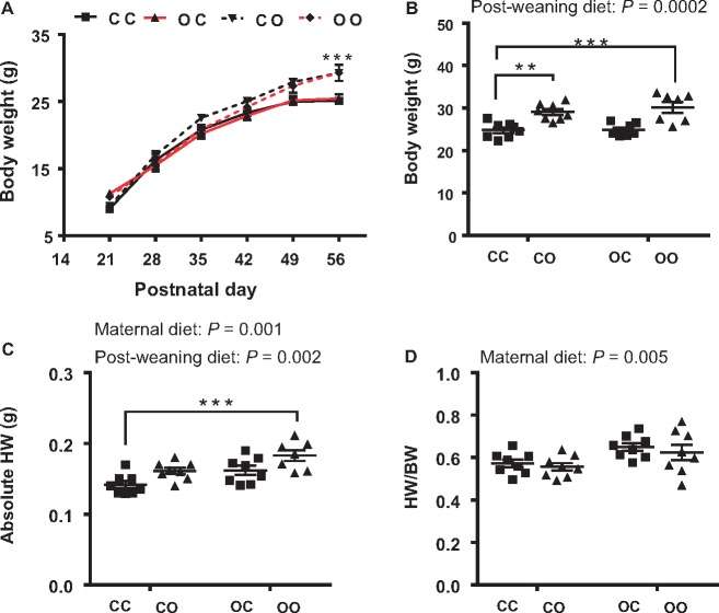
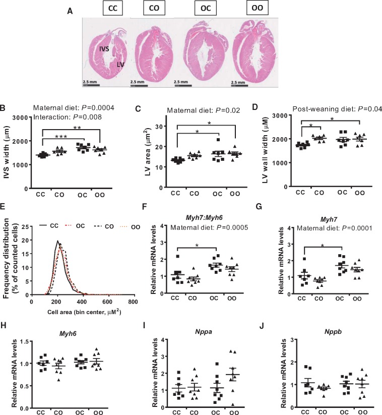
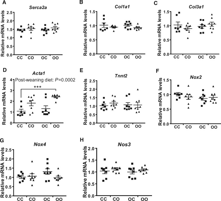
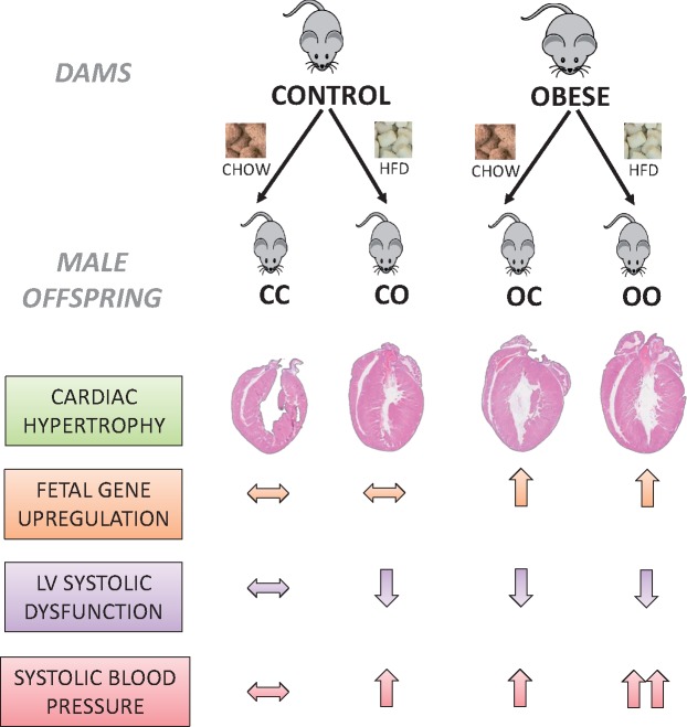
Methods and results: The impact of the maternal and postnatal environment on the offspring metabolic profile, arterial blood pressure, cardiac structure, and function was assessed in 8-week-old C57BL/6 male mice. Measurement of cardiomyocyte cell area, the transcriptional re-activation of cardiac fetal genes as well as genes involved in the regulation of contractile function and matrix remodelling in the adult heart were determined as potential mediators of effects on cardiac function. In the adult offspring: a post-weaning obesogenic diet coupled with exposure to maternal obesity increased serum insulin (P < 0.0001) and leptin levels (P < 0.0001); maternal obesity (P = 0.001) and a post-weaning obesogenic diet (P = 0.002) increased absolute heart weight; maternal obesity (P = 0.01) and offspring obesity (P = 0.01) caused cardiac dysfunction but effects were not additive; cardiac dysfunction resulting from maternal obesity was associated with re-expression of cardiac fetal genes (Myh7: Myh6 ratio; P = 0.0004), however, these genes were not affected by offspring diet; maternal obesity (P = 0.02); and offspring obesity (P = 0.05) caused hypertension and effects were additive.

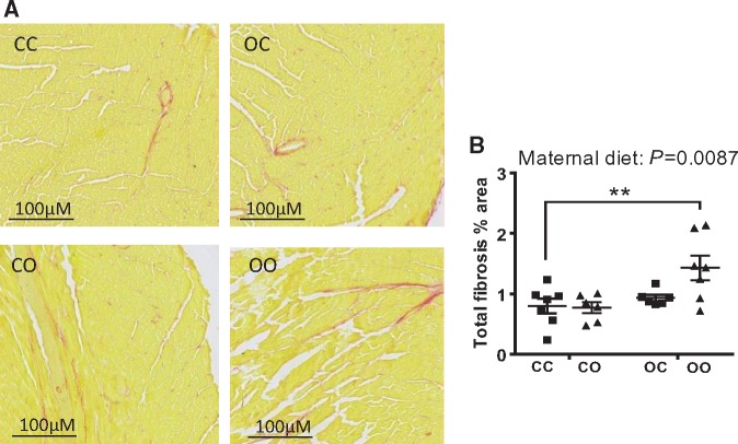
Conclusions: Maternal diet-induced obesity and offspring obesity independently promote cardiac dysfunction and hypertension in adult male progeny. Exposure to maternal obesity alone programmed cardiac dysfunction, associated with hallmarks of pathological left ventricular hypertrophy, including increased cardiomyocyte area, upregulation of fetal genes, and remodelling of cardiac structure. These data highlight that the perinatal period is just as important as adult-onset obesity in predicting CVD risk. Therefore, early developmental periods are key intervention windows to reduce the prevalence of CVD.


Figures

Figure 1
Figure 1
Exposure to maternal over-nutrition is sufficient to increase heart weight. (A) Offspring growth curve from weaning to 8 weeks (in red are groups exposed to maternal obesity); (B) body weight at 8 weeks of age; (C) absolute heart weight; (D) heart weight normalized to body weight. N = 7–8 male mice from different dams were used for each group. CC, mum fed chow, offspring fed chow; OC, mum fed obesogenic diet, offspring fed chow; CO, mum fed chow, offspring fed obesogenic diet; OO, mum fed obesogenic diet, offspring fed obesogenic diet. Two-way ANOVA followed by Dunnet’s post hoc test for multiple comparisons vs. CC was performed. *P < 0.05, **P < 0.01, ***P < 0.001.
Figure 2
Figure 2
Exposure to maternal over-nutrition programmes pathological cardiac hypertrophy. (A) Representative H&E staining of mid-cardiac sections; (B) interventricular septum (IVS) width; (C) area of the left ventricle; (D) left ventricular (LV) wall width. N= 6–7 animals from independent litters for each dietary group. (E) Frequency distribution of cardiomyocytes cell area in the left ventricle of CC, OC, CO, and OO 8-week-old animals (in red are groups exposed to maternal obesity; n = 6/dietary group); (F–J) mRNA levels of cardiac hypertrophic markers Myh7: Myh6, Myh7, Myh6, Nppa, and Nppb (n = 7–8). CC, mum fed chow, offspring fed chow; OC, mum fed obesogenic diet, offspring fed chow; CO, mum fed chow, offspring fed obesogenic diet; OO, mum fed obesogenic diet, offspring fed obesogenic diet. Two-way ANOVA followed by Dunnett’s post hoc test for multiple comparisons vs. CC was performed. The overall effect of maternal diet and post-weaning diet is reported where significant in each panel. Significant vs. CC: *P < 0.05, **P < 0.01, ***P < 0.001.
Figure 3
Figure 3
Male offspring exposed to maternal obesity develop signs of cardiac fibrosis. (A) Representative images of ventricular mid-cardiac sections (10 µm) stained with Picrosirius Red. Areas positive for fibrosis were stained red; (B) quantification of total fibrosis expressed as a percentage of tissue area. CC, mum fed chow, offspring fed chow; OC, mum fed obesogenic diet, offspring fed chow; CO, mum fed chow, offspring fed obesogenic diet; OO, mum fed obesogenic diet, offspring fed obesogenic diet. Data are presented as means ± SEM (n = 6–8 from independent litters for each dietary group) and were analysed by two-way ANOVA. Dunnett’s multiple comparisons test was performed to evaluate significant differences to the CC group: **P < 0.01.
Figure 4
Figure 4
Maternal obesity only partially affects the expression of contractile and matrix remodelling genes in offspring hearts. Gene expression of collagen encoding for Serca2a (A), Col1a1 (B), Col3a1 (C), Acta1 (D), Tnnt2 (E), Nox2 (F), Nox4 (G), and Nos3 (H) in the cardiac tissue of the four experimental groups, expressed relative to CC group. CC, mum fed chow, offspring fed chow; OC, mum fed obesogenic diet, offspring fed chow; CO, mum fed chow, offspring fed obesogenic diet; OO, mum fed obesogenic diet, offspring fed obesogenic diet. Data are presented as means ± SEM (n = 7–8 from independent litters for each dietary group) and were analysed by two-way ANOVA and overall effect of post-weaning diet is reported where significant. Dunnett’s post hoc test for multiple comparisons test was then performed to evaluate significant differences to the CC group: ***P < 0.001.
Figure 5
Figure 5
Schematic diagram of the findings of this study showing distinct programming mechanisms of hypertension and cardiac dysfunction by maternal diet-induced obesity.

Comment in

References

    1. Lauby-Secretan B, Scoccianti C, Loomis D, Grosse Y, Bianchini F, Straif K, Group IAfRoCHW. Body fatness and cancer–viewpoint of the IARC Working Group. N Engl J Med 2016;375:794–798. - PMC - PubMed
    1. Townsend N, Wilson L, Bhatnagar P, Wickramasinghe K, Rayner M, Nichols M.. Cardiovascular disease in Europe: epidemiological update 2016. Eur H J 2016;37:3232–3245. - PubMed
    1. Poston L, Caleyachetty R, Cnattingius S, Corvalán C, Uauy R, Herring S, Gillman MW.. Preconceptional and maternal obesity: epidemiology and health consequences. Lancet Diabetes Endocrinol 2016;4:1025–1036. - PubMed
    1. Zambrano E, Ibáñez C, Martínez-Samayoa PM, Lomas-Soria C, Durand-Carbajal M, Rodríguez-González GL.. Maternal obesity: lifelong metabolic outcomes for offspring from poor developmental trajectories during the perinatal period. Arch Med Res 2016;47:1–12. - PubMed
    1. Gluckman PD, Hanson MA, Cooper C, Thornburg KL.. Effect of in utero and early-life conditions on adult health and disease. N Engl J Med 2008;359:61–73. - PMC - PubMed

Publication types

MeSH terms