. 2019 Jan 1;16(1):59-70.
doi: 10.1093/ons/opy034.

Folate Receptor Near-Infrared Optical Imaging Provides Sensitive and Specific Intraoperative Visualization of Nonfunctional Pituitary Adenomas

Affiliations

Folate Receptor Near-Infrared Optical Imaging Provides Sensitive and Specific Intraoperative Visualization of Nonfunctional Pituitary Adenomas

Steve S Cho et al. Oper Neurosurg. .

Abstract

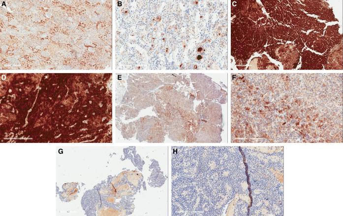
Background: Surgical resection is the primary treatment for nonfunctional (NF) pituitary adenomas, but gross-total resection is difficult to achieve in all cases. NF adenomas overexpress folate receptor alpha (FRα).

Objective: To test the hypothesis that we could target FRα for highly sensitive and specific intraoperative detection of NF adenomas using near-infrared (NIR) imaging.

Methods: Fourteen patients with NF pituitary adenoma were infused with the folate analog NIR dye OTL38 preoperatively. NIR fluorescence signal-to-background ratio (SBR) was recorded for each tumor during resection of the adenomas. Extent of surgery was not modified based on the presence or absence of fluorescence. Immunohistochemistry was performed to assess FRα expression in all specimens. Magnetic resonance imaging (MRI) was performed postoperatively to assess residual neoplasm.

Results: Nine adenomas overexpressed FRα and fluoresced with a NIR SBR of 3.2 ± 0.52, whereas the 5 non-FRα-overexpressing adenomas fluoresced with an SBR of 1.5 ± 0.21. Linear regression demonstrated a significant correlation between intraoperative SBR and the FRα expression (P-value < .001). Analysis of 14 margin samples revealed that the surgeon's impression of the tissue had 83% sensitivity, 100% specificity, 100% positive predictive value, and 89% negative predictive value, while NIR fluorescence had 100% for all values. NIR fluorescence accurately predicted postoperative MRI results in 78% of FRα-overexpressing patients.

Conclusion: Preoperative injection of folate-tagged NIR dye provides strong signal and visualization of NF pituitary adenomas. It is 100% sensitive and specific for detecting margin neoplasm and can predict postoperative MRI findings. Our results suggest that NIR fluorescence may be superior to white-light visualization alone and may improve resection rates in NF pituitary adenomas.


Figures

FIGURE 1.
FIGURE 1.
Subject 31 with FRα H-score of 250 demonstrates strong NIR fluorescence and GTR is achieved by removing fluorescent specimen. A and B, Sagittal and coronal preoperative MRI demonstrating a 20-mm sellar mass with a Knosp grade 2 invasion of the cavernous sinus. C and D, Upon initial exposure after dura opening, the tumor is visualized with white light in C, and it can be seen in real time with NIR overlay with an SBR of 3.1 in D. E and F, White light alone does not show convincing residual E, but the NIR overlay view demonstrates a piece of inferior dura with positive NIR signal with an SBR of 2.5 (F, white circle). This sample was believed to be dura only with no neoplasm and was taken in entirety as a margin sample by the senior surgeon (J.Y.K.L.). However, pathology revealed that portions of the specimen were neoplastic. G and H, Day 1 postoperative sagittal and coronal MRI does not demonstrate any evidence of residual neoplasm.
FIGURE 2.
FIGURE 2.
Subject 26 with FRα H-score of 200 demonstrates strong NIR fluorescence, with residual neoplasm. A and B, Sagittal and coronal preoperative MRI demonstrating a 27-mm sellar mass with a Knosp grade 2 invasion of the cavernous sinus. C and D, Upon initial exposure after the dura has been opened, the tumor can be seen with the white light view in C, and it can be seen in real time with NIR overlay directly on top of the white light view in D, with an SBR of 3.9. E and F, White light does not show convincing residual E, but the NIR overlay view shows strong residual fluorescence with an SBR of 3.4 protruding on the left anterior side of the sella. This margin specimen was not biopsied by the senior surgeon (M.S.G.). G and H, Two-month postoperative sagittal and coronal MRI demonstrate evidence of residual tumor in the anterior part of the sella (white arrows), consistent with the fluorescent area seen in F.
FIGURE 3.
FIGURE 3.
Immunohistochemistry demonstrates level of FRα expression in pituitary adenomas. A, Kidney as positive control with H-score of 190 (scale bar = 600 μm). B, Normal adenohypophysis with H-score 34 (scale bar = 200 μm). C and D, Low and high power view (scale bars = 600, 200 μm respectively) of a null-cell adenoma subject ID 12 with high H-score of 270. E and F, Low and high power view (scale bars = 600, 200 μm respectively) of somatotroph adenoma of subject ID 2 with intermediate H-score of 100. G and H, Low and high power view (scale bars = 600, 200 μm respectively) of a null-cell adenoma of subject ID 5 with low H-score of 0. The red areas in panels G and H are red blood cells and folate staining was only measured in the blue areas, where the pituitary cells are present.
FIGURE 4.
FIGURE 4.
Intraoperative NIR fluorescence SBR strongly correlates to FRα expression levels. The 9 adenomas with FRα H-scores ≥ 200 demonstrate mean SBR of 3.2 ± 0.52, while the 5 adenomas with FRα H-scores ≤ 100 demonstrate mean SBR of 1.5 ± 0.18 (P-value < .001). At the time of surgery, only the NIR SBR is known. Thus, being able to predict FRα expression levels during surgery is crucial. This linear regression analysis (95% CI demonstrated in shaded area) demonstrates significant correlation between intraoperative SBR and FRα expression (P-value < .001). Each 1-point increase in SBR correlates to approximately 108-point change in FRα H-score.
FIGURE 5.
FIGURE 5.
Limitations of OTL38 and linear endoscope in detecting residual neoplasm in the cavernous sinus. NIR imaging still requires direct line of sight which cannot always be accomplished with the VisionSense zero-degree endoscope. A and B, Preoperative sagittal and coronal MRI in subject 21 with FRα H-score of 260 demonstrate a 20-mm adenoma with a Knosp grade 4 invasion of the left cavernous sinus. C and D, Upon initial exposure after dura opening, the tumor can be seen with both white light alone C and the NIR-overlay view with a NIR SBR of 3.4 D. E and F, Upon completion of surgery using white light, the NIR camera is introduced to assess for margins. The NIR-overlay view shows mild areas of NIR fluorescence (SBR = 2.5) in the left inferior sella F. This area was not biopsied by the senior surgeon (M.S.G.), but it corresponds to the 2-mo follow-up sagittal MRI showing sellar residual (G, white arrow). The same view, however, does not show the left cavernous sinus neoplasm surrounding the internal carotid artery, seen in the 2-mo follow-up coronal MRI (H, white arrow). Thus, a major limitation of the current technique is the limited views around corners into the cavernous sinus with the zero-degree endoscope. The patient required follow-up Gamma Knife stereotactic surgery (Elekta AB) 1 yr after the surgery.

References

    1. Ezzat S, Asa SL, Couldwell WTet al.. The prevalence of pituitary adenomas. Cancer. 2004;101(3):613-619. - PubMed
    1. Greenman Y, Cooper O, Yaish Iet al.. Treatment of clinically nonfunctioning pituitary adenomas with dopamine agonists. Eur J Endocrinol. 2016;175(1):63-72. - PubMed
    1. Lee JYK, Bohman L-E, Bergsneider M. Contemporary neurosurgical techniques for pituitary tumor resection. J Neurooncol. 2014;117(3):437-444. - PubMed
    1. Losa M, Mortini P, Barzaghi Ret al.. Early results of surgery in patients with nonfunctioning pituitary adenoma and analysis of the risk of tumor recurrence. J Neurosurg. 2008;108(3):525-532. - PubMed
    1. Thawani J, Ramayya A, Pisapia J, Abdullah K, Lee J, Grady M. Operative strategies to minimize complications following resection of pituitary macroadenomas. J Neurol Surg Part B Skull Base. 2017;78(2):184-190 - PMC - PubMed

Publication types

Substances